Bone scan
Peer reviewed by Dr Colin Tidy, MRCGPLast updated by Dr Doug McKechnie, MRCGPLast updated 13 Mar 2023
Meets Patient’s editorial guidelines
- DownloadDownload
- Share
- Language
- Discussion
- Audio Version
- Add to preferred sources on Google
A bone scan is a special type of nuclear medicine procedure that uses radionuclides to create a picture of the bones. Radionuclides are chemicals which emit radioactivity that can be detected by special scanners.
A bone scan is different to a bone density scan (DEXA). See the DEXA Scan leaflet for more information on this bone density test. A DEXA scan is also known as a DXA scan.
Note: the information below is a general guide only. The arrangements, and the way tests are performed, may vary between different hospitals. Always follow the instructions given by your doctor or local hospital.
At a glance
A bone scan uses a small amount of radioactive fluid to create pictures of your bones.
It can help detect cancer, infections, broken bones, and other causes of bone pain.
You will receive an injection and wait a few hours before the scan takes place.
The scan itself typically takes between 30 minutes and 1.5 hours.
There is a small risk to unborn babies, so tell your doctor if you are pregnant or think you might be.
You should avoid close contact with children or pregnant women for 24 hours after the scan.
In this article:
Video picks for Imaging
Continue reading below
How does a bone scan work?
Bone scans use radionuclides to detect areas of the bone which are growing or being repaired. A radionuclide (sometimes called a radioisotope or isotope) is a chemical which emits a type of radioactivity called gamma rays. A tiny amount of radionuclide is put into the body, usually by an injection into a vein.
Cells which are most 'active' in the target tissue or organ will take up more of the radionuclide. So, active parts of the tissue will emit more gamma rays than less active or inactive parts.
Gamma rays are similar to X-rays and are detected by a device called a gamma camera. The gamma rays which are emitted from inside the body are detected by the gamma camera. The rays are then converted into an electrical signal and sent to a computer.
The computer builds a picture by converting the differing intensities of radioactivity emitted into different colours or shades of grey. For example, areas of the target organ or tissue which emit lots of gamma rays may be shown as red spots ('hot spots') on the picture on the computer monitor.
Areas which emit low levels of gamma rays may be shown as blue ('cold spots'). Various other colours may be used for 'in between' levels of gamma rays emitted.
How long does a bone scan usually take?
In total, you should allow about four to six hours for everything required for the scan.
An injection of radionucleotides is given via a drip (cannula) into your vein. This takes about 2 to 4 hours to circulate around the body. Sometimes, a few scans might be taken straightaway. You will have the main part of the bone scan after a waiting period of a few hours.
Once enough time has passed, you'll have the main scan. This can take between 30 minutes and 1.5 hours; the radiographers doing the scan will be able to tell you how long to expect.
Are bone scans safe?
Back to contentsThe term 'radioactivity' may sound alarming. But, the radioactive chemicals used in radionuclide scans are considered to be safe, and they leave the body quickly in the urine. The amount of radiation that your body receives is very small. In many cases, the level of radiation involved is not much different to a series of a few normal X-rays. However:
As with any other types of radiation (such as X-ray), there is a small risk that the gamma rays may affect an unborn child. So, tell your doctor if you are pregnant or if you may be pregnant.
It takes about 24 hours for the radionucleotide to leave your body completely. The risk to other people is very, very low (and might even be zero), but, as a precaution, you'll often be advised to avoid prolonged close contact with children or pregnant women for 24 hours.
Rarely, some people have had allergic reactions to the injected chemical. Tell your doctor if you are allergic to iodine.
Theoretically, it is possible to receive an overdose when the chemical is injected. This is very rare.
Continue reading below
What does a bone scan show?
Back to contentsIn a bone scan, a radionuclide is used which collects in areas where there is a lot of bone activity (where bone cells are breaking down or repairing parts of the bone).
So a bone scan is used to detect:
Cancer that has spread to the bones.
Infection of the bone, including infections around joint replacements.
Broken bones that can't be seen on other tests.
Bone damage.
Other causes of unexplained bone pain.
These areas of activity are seen as 'hot spots' on the scan picture.
This type of radionuclide bone scanning is also called bone scintigraphy. It is a totally different type of procedure to the DEXA bone scan which is used to measure density of bones in conditions such as 'thinning' of the bones (osteoporosis). (See the separate leaflet called DEXA Scan for information about this other kind of bone scan.)
What preparation do I need?
Back to contentsUsually very little. Your hospital should provide you with information regarding any special arrangements. This test should not be carried out in pregnant women. You should advise your doctor if you are pregnant or, if you think you may be pregnant.
You should also inform your hospital if you are breastfeeding, as special precautions may be necessary. You may also be asked to empty your bladder of urine before the scanning begins. You will be asked to drink several glasses of water between the injection and the scan.
Continue reading below
What happens during a bone scan?
Back to contentsWhen you arrive, a radiographer (a specially trained person who operates the radiology machines to get the scan images) will explain what will happen, and will ask you to sign a consent form to indicate that you understand and agree to go ahead. You can, and should, ask any questions you have about the procedure at this point.
The injection
In a bone scan a small amount of a radioactive substance (radionuclide tracer) is injected into a vein in your arm. It then takes some time after injection of the tracer - sometimes several hours - for the radionuclide to travel to the target tissue and to be 'taken' into the active cells. So, after receiving the radionuclide you may have a wait of a few hours. You may be able to go out and come back to the scanning room later in the day.
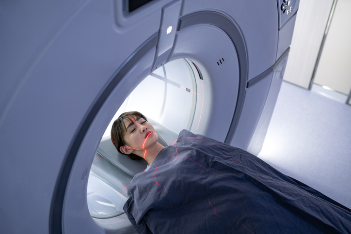
The scan
When it is time to do the scanning, you will need to lie on a couch while the gamma camera detects the gamma rays coming from your body, and the computer turns the information into a picture. You need to lie as still as possible whilst each picture is taken (so it is not blurred). Some pictures can take 30 minutes or more.
The number of pictures taken, and the time interval between each picture, vary depending on what is being scanned. For a whole body bone scan, you move slowly through the whole scanner and the picture is taken continuously.
After the bone scan
Bone scans do not generally cause any after effects. Through the natural process of radioactive decay, the small amount of radioactive chemical in your body will lose its radioactivity over time.
It also passes out of your body through your urine over about 24 hours. You may be instructed to take special precautions after urinating, to flush the toilet twice and to wash your hands thoroughly.
You will be advised to drink plenty of water for a day after the scan to help flush the radionuclide out of your system.
If you have contact with children or pregnant women you should let your doctor know. Although the levels of radiation used in the scan are small they may advise special precautions. Your hospital should give you more advice on this.
Patient picks for Imaging
Tests and investigations
Radionuclide scan
A radionuclide scan is a way of imaging bones, organs and other parts of the body by using a small dose of a radioactive chemical. There are different types of radionuclide chemical. The one used depends on which organ or part of the body is to be scanned. Note: the information below is a general guide only. The arrangements, and the way tests are performed, may vary between different hospitals. Always follow the instructions given by your doctor or local hospital.
by Dr Rachel Hudson, MRCGP
Tests and investigations
MRI scan
An MRI scan is a safe and painless test that can provide detailed pictures of organs and other structures inside your body. Note: the information below is a general guide only. The arrangements (and the way tests are performed) may vary between different hospitals. Always follow the instructions given by your doctor or local hospital. These are usually included with your appointment letter.
by Dr Toni Hazell, MRCGP
Frequently asked questions
What is a radionuclide and how does it work in a bone scan?
A radionuclide is a chemical that gives off gamma rays, which are a type of radioactivity similar to X-rays. A small amount is injected into your body, usually into a vein. These radionuclides are then absorbed more readily by areas of bone that are highly active, for example, where new bone is growing or being repaired. The gamma rays emitted from these active areas are detected by a special camera, which then creates an image on a computer showing these 'hot spots'.
Why do I have to wait several hours after the injection before the main scan?
After the radionuclide is injected, it needs time to circulate throughout your body and be taken up by the active cells in your bones. This process can take several hours, typically between 2 to 4 hours, to ensure enough of the substance has collected in the target areas before the gamma camera can accurately detect it and create a clear image.
What happens if I'm not able to lie still during the scan?
It is important to lie as still as possible during the scan, as movement can blur the pictures captured by the gamma camera. Some pictures can take a significant amount of time, up to 30 minutes or more, and continuous movement during these times could affect the clarity and usefulness of the scan results.
What should I do after the scan to help remove the radioactive substance from my body?
After the scan, it is recommended to drink plenty of water for about a day. This helps your body flush out the radionuclide through your urine. You may also be advised to take specific precautions after urinating, such as flushing the toilet twice and washing your hands thoroughly, to minimise any residual radioactivity.
Can I go home between the injection and the main scan?
Yes, after receiving the radionuclide injection, there is a waiting period of a few hours for the substance to travel to the target tissues. Depending on the hospital's policy and the specific procedure, you may be able to leave and return to the scanning room later in the day for the main part of your scan.
Further reading and references
- O'Sullivan GJ, Carty FL, Cronin CG; Imaging of bone metastasis: An update. World J Radiol. 2015 Aug 28;7(8):202-11. doi: 10.4329/wjr.v7.i8.202.
- Graham R, Little D, Cade S, et al; British Nuclear Medicine Society Clinical Guideline for bone scintigraphy. Nucl Med Commun. 2022 Nov 1;43(11):1109-1112. doi: 10.1097/MNM.0000000000001615. Epub 2022 Sep 27.
Continue reading below
About the authorView full bio

Dr Doug McKechnie, MRCGP
Medical Writer
MA, MBBS, MSc, DRCOG, MRCP(UK), MRCGP(2021), FHEA
Dr Doug McKechnie is an NHS GP working in London. He works full-time clinically and is also the Deputy Lead for the Clinical and Professional Practice module at University College London Medical School.
About the reviewerView full bio

Dr Colin Tidy, MRCGP
General Practitioner, Medical Author
MBBS, MRCGP, MRCP (Paediatrics), DCH
Dr Colin Tidy is an NHS Doctor, based in Oxfordshire.
Article history
The information on this page is written and peer reviewed by qualified clinicians.
Next review due: 11 Mar 2028
13 Mar 2023 | Latest version

Ask, share, connect.
Browse discussions, ask questions, and share experiences across hundreds of health topics.

Feeling unwell?
Assess your symptoms online for free
Sign up to the Patient newsletter
Your weekly dose of clear, trustworthy health advice - written to help you feel informed, confident and in control.
By subscribing you accept our Privacy Policy. You can unsubscribe at any time. We never sell your data.