Pleuraerguss
Begutachtet von Dr Doug McKechnie, MRCGPZuletzt aktualisiert von Dr Hayley Willacy, FRCGP Last updated 21 Dec 2023
Erfüllt die Anforderungen des Patienten Richtlinien des Patienten
- HerunterladenHerunterladen
- Teilen
- Language
- Diskussion
- Audio-Version
- Add to preferred sources on Google
Ein Pleuraerguss ist eine Flüssigkeitsansammlung neben der Lunge. Es gibt verschiedene Ursachen. Der Erguss kann dazu führen, dass Sie Atemnot bekommen. Die Flüssigkeit kann bei Bedarf abgeleitet werden. Die Behandlung zielt hauptsächlich auf die zugrunde liegende Ursache ab.
At a glance
A pleural effusion is a build-up of fluid between a lung and the chest wall.
This fluid build-up can separate the lung from the chest wall.
Common causes include infections, heart failure, and some cancers.
Symptoms can include breathlessness and, sometimes, chest pain.
A chest X-ray usually confirms a pleural effusion.
Treatment often involves addressing the underlying cause.
In diesem Artikel:
Video picks for Brust- und Lungenerkrankungen
Lesen Sie unten weiter
What is a pleural effusion?
Lunge und Atemwege mit Pleura
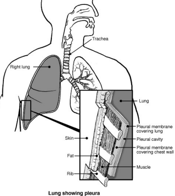
A pleural effusion means that there is a build-up of fluid in the space between a lung and the chest wall.
The pleura is a thin membrane that lines the inside of the chest wall and covers the lungs. There is normally a tiny amount of fluid between the two layers of pleura. This acts like lubricating oil between the lungs and the chest wall as they move when you breathe.
A pleural effusion develops when this fluid builds up and separates the lung from the chest wall.
Types of pleural effusions
Zurück zum InhaltPleural effusions are classed as transudates or exudates according to how much protein they contain.
Transudates have a low protein level of <25g/L. Fluid accumulates due to a disruption in the balance of fluid pressures.
Exudates have a high protein level of >35g/L. Fluid accumulates due to increased leakiness (permeability) of the smallest blood vessels.
Transudative pleural effusions
These are most often caused by conditions such as heart failure and liver failure. Less common causes are nephrotisches Syndrom, Hypothyreose and as a consequence of peritoneal dialysis.
Exudative pleural effusions
The most common causes of these are infections, such as Lungenentzündung oder Tuberkulose, or cancer. Less common causes include autoimmune conditions such as rheumatoide Arthritis, after a Herzinfarkt und Pankreatitis.
Lesen Sie unten weiter
Causes of a pleural effusion
Zurück zum InhaltA pleural effusion is a complication of various conditions. The following are some of the more common causes of a pleural effusion (but there are other rarer causes too):
Lung infection (pneumonia), Tuberkulose, and cancers may cause inflammation of the lung and pleura. This may cause fluid to build up into a pleural effusion.
Some arthritic conditions may cause inflammation of the pleura in addition to joint inflammation. For example, pleural effusion is an uncommon complication of rheumatoide Arthritis und systemischer Lupus erythematodes (SLE).
Herzinsuffizienz causes 'back pressure' in the veins (blood vessels) that take blood back to the heart. Some fluid may seep out of the blood vessels. Swelling of the legs with fluid is typical with heart failure, but a pleural effusion may also develop.
A low level of protein in the blood also tends to allow fluid to seep out of the blood vessels. For example, cirrhosis of the liver and some kidney diseases may cause a low level of blood protein which allows a pleural effusion to develop.
Symptoms of a pleural effusion
Zurück zum InhaltYou may feel some chest pain but a pleural effusion is often painless. The amount of fluid varies. As the effusion becomes larger, it presses on the lung, which cannot expand fully when you breathe. You may then become breathless.
You may also have symptoms of the condition that is causing the effusion. As a whole range of conditions can cause a pleural effusion, there is a large range of other symptoms that may occur, depending on the underlying cause. One example is you may have a cough and a high temperature (fever) if the cause is lung infection (pneumonia).
Lesen Sie unten weiter
How is a pleural effusion diagnosed?
Zurück zum InhaltA Röntgenbild des Brustkorbs usually confirms a build-up of fluid between a lung and the chest wall (pleural effusion). If the cause of the effusion is known then no further tests may be needed. However, sometimes a pleural effusion is the first sign of an underlying condition.
Further tests may then be advised to find the cause of the effusion. These may include lung tests, Bluttests and taking a sample of the fluid and pleura to examine in the laboratory.
Pleural effusion treatment
Zurück zum InhaltTreating the underlying cause
A major part of treatment is usually directed to the underlying cause of the build-up of fluid between the lung and the chest wall (pleural effusion). For example, medicines called Antibiotika for lung infection (pneumonia), Chemotherapie oder Strahlentherapie for cancers, etc.
Therefore, treatment can vary greatly, depending on the cause of the effusion. If the underlying cause can be successfully treated then there is a good chance that the pleural effusion will go away for good. If the underlying cause cannot be treated, or can only be partially treated, the effusion may return if it is cleared (drained).
Treating the effusion itself
Small effusions that cause no symptoms, or only mild symptoms, may just be left and 'observed'. Treatment is usually only needed if the effusion causes symptoms such as breathlessness.
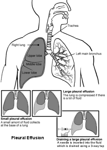
A large pleural effusion that makes you breathless can be drained. This is called a pleural fluid aspiration or pleural tap. It is usually done by inserting a needle or tube through the chest wall. A local anaesthetic is injected into the skin and chest wall first to make the procedure painless. This may be a 'one-off' procedure to relieve symptoms.
Lungs and airways - pleural effusion
However, in many cases, unless the underlying cause can be treated, an effusion is likely to return within a few weeks. Repeated draining of the fluid, when symptoms become troublesome, is one option.
Depending on the underlying cause, other treatment options that are sometimes considered include:
Pleurodesis.
In this procedure, a special chemical (a sclerosant) is injected into the pleural space. This causes inflammation of the pleural membranes and helps them to 'stick' together. This helps to prevent fluid building up again into an effusion. Pleurodesis is most often used in the treatment of repeated (recurrent) effusions caused by cancer.
Talc pleurodesis is often used. If the lung re-inflates after the fluid has been drained, sterile talcum powder (talc) can be used to help stick the pleura together. The doctor puts the talc through the tube attached to the drain and then leaves the drain clamped for about an hour. This allows time for the 2 linings of the lung to stick together. The doctor may attach the drain to a suction machine to apply a small amount of pressure. This can help the pleura to seal together. After a pleurodesis, you will usually have the drain in place for another 24 hours.
Permanent drains
If the effusion is recurrent (perhaps because the underlying cause is not treatable), it is possible to have repeated chest drains put in. This may be uncomfortable and mean spending a lot of time going in and out of the hospital. In this situation, a special catheter called a tunnelled indwelling pleural catheter (TIPC), can be put in. This allows the pleural effusion to be drained easily while you are at home. When you feel you have a build-up of fluid on the lung, the catheter allows the fluid to be drained off into a bottle. This can be done by you, a family member or a nurse. The catheter is inserted in hospital on a day ward. As long as there are no complications, you can usually go home on the same day.
Shunt insertion
An operation to insert a shunt (like an internal drain) to allow the fluid to drain out from the chest into the tummy (abdominal) cavity. This is called a 'pleuroperitoneal shunt'. It is only occasionally used.
Video assisted thorascopic surgery
It may be possible to drain a pleural effusion and do a pleurodesis using a procedure called a thoracoscopy. This is a type of keyhole surgery where the doctor puts a small flexible tube (a thorascope) into the chest. The tube has a light and camera at the end, so the doctors can see into your chest. This is why it is called 'video assisted'. It can be used for taking a biopsy, for draining an effusion or pleurodesis. For this procedure, you lie on your side and are given an injection of a sedative to make you feel drowsy. Local anaesthetic is used to numb the area and the doctor makes a small cut to put the thorascope in. The procedure takes about 40 to 60 minutes and afterwards a chest drain will be left to drain any remaining fluid from the chest cavity.
Surgery also includes an operation to remove the pleura called a pleurectomy. It is sometimes used in people with effusions due to cancer when other treatment options have failed.
Patient picks for Brust- und Lungenerkrankungen

Brust und Lungen
Pulmonale Fibrose
Lungenfibrose ist eine schwere Lungenerkrankung, bei der die kleinen Luftsäcke der Lunge (Alveolen) und das Lungengewebe neben den Alveolen beschädigt und vernarbt werden, was zu Lungenfibrose führt. Das Hauptsymptom ist Atemnot, die allmählich schlimmer wird. Die Ursache ist oft unbekannt, aber Lungenfibrose kann durch verschiedene zugrunde liegende Erkrankungen verursacht werden. Die Behandlung umfasst die Behandlung der zugrunde liegenden Ursache sowie die Verabreichung von Steroiden und anderen Medikamenten. Immer häufiger wird eine Lungentransplantation in Betracht gezogen.
von Dr. Toni Hazell, MRCGP

Brust und Lungen
Bronchiektasie
Bronchiektasie ist ein Problem mit den Lungen, bei dem Sie viel Schleim (Sputum) abhusten: deutlich mehr als üblich. Es wird meist durch eine bereits die Lunge beeinträchtigende Ursache verursacht, wie eine schwere Infektion; manchmal wird jedoch keine Ursache gefunden. Es betrifft in der Regel ältere Menschen. Es gibt einige gute Behandlungsmöglichkeiten, um die Erkrankung unter Kontrolle zu halten.
by Dr Hayley Willacy, FRCGP
Häufig gestellte Fragen
What is the pleura and how does it relate to a pleural effusion?
The pleura is a thin membrane that lines the inside of your chest wall and covers your lungs. Normally, there's a small amount of fluid between these two layers, which acts like a lubricant when your lungs move during breathing. A pleural effusion occurs when too much of this fluid builds up, separating the lung from the chest wall.
Are there different kinds of pleural effusions?
Yes, pleural effusions are classified into two main types based on their protein content: transudates and exudates. Transudates have low protein levels and usually result from an imbalance in fluid pressures, often linked to conditions like heart or liver failure. Exudates have high protein levels and are typically caused by increased leakiness of blood vessels, commonly due to infections or cancer.
If I have a pleural effusion, will I always feel chest pain?
Not necessarily. While some people may experience chest pain, a pleural effusion is often painless. Symptoms like breathlessness usually develop as the effusion becomes larger, because the fluid presses on the lung, preventing it from expanding fully when you breathe.
How quickly can a pleural effusion return after being drained?
If the underlying cause of the pleural effusion cannot be treated, or only partially treated, the fluid is likely to return within a few weeks after being drained. In such cases, your doctor might discuss options like repeated draining, pleurodesis, or permanent drains to manage recurrent effusions.
What is pleurodesis and why might it be recommended?
Pleurodesis is a procedure where a special chemical, like talc, is injected into the space between the lung and chest wall. This causes the pleural membranes to become inflamed and stick together, which helps prevent fluid from building up again. It is most often used for repeated pleural effusions, especially those caused by cancer, after the fluid has been drained.
What is a tunnelled indwelling pleural catheter (TIPC) and how is it used?
A TIPC is a special catheter that can be inserted into the chest to allow fluid to be drained easily at home when a pleural effusion is recurrent. This avoids repeated hospital visits for drainage. You, a family member, or a nurse can drain the fluid into a bottle when you feel a build-up. The catheter is inserted in the hospital, usually as a day procedure.
Can surgery cure a pleural effusion?
Surgery can be an option for managing pleural effusions, especially recurrent ones or those caused by cancer. Procedures like thoracoscopy can drain the fluid and perform a pleurodesis. In some cases where other treatments haven't worked, an operation called a pleurectomy, which removes the pleura, might be used. These surgical options aim to either manage the fluid build-up or address the underlying cause locally.
Weiterführende Literatur und Referenzen
- Leitlinie der British Thoracic Society für Pleuraderkrankungen; British Thoracic Society - BMJ (2023).
- Krishna R, Rudrappa M; Pleural Effusion. StatPearls 2020.
- Skok K, Hladnik G, Grm A, et al; Malignant Pleural Effusion and Its Current Management: A Review. Medicina (Kaunas). 2019 Aug 15;55(8). pii: medicina55080490. doi: 10.3390/medicina55080490.
- Bibby AC, Dorn P, Psallidas I, et al; ERS/EACTS statement on the management of malignant pleural effusions. Eur Respir J. 2018 Jul 27;52(1). pii: 13993003.00349-2018. doi: 10.1183/13993003.00349-2018. Print 2018 Jul.
- Kulandaisamy PC, Kulandaisamy S, Kramer D, et al; Malignant Pleural Effusions-A Review of Current Guidelines and Practices. J Clin Med. 2021 Nov 26;10(23). pii: jcm10235535. doi: 10.3390/jcm10235535.
Lesen Sie unten weiter
About the authorView full bio

Dr Hayley Willacy, FRCGP
Allgemeinmediziner, Medizinischer Autor
MBChB (1992), DRCOG, DFFP, MRCOG (Part 1) MRCGP (2007), DFSRH (2013), MSc - medical education (2020)
Dr Hayley Willacy was an NHS GP working in northwest England, who retired from clinical practice in 2022 after 30 years.
About the reviewerView full bio

Dr Doug McKechnie, MRCGP
Medizinischer Autor
MA, MBBS, MSc, DRCOG, MRCP(UK), MRCGP(2021), FHEA
Dr. Doug McKechnie ist ein NHS-Hausarzt, der in London arbeitet. Er arbeitet klinisch in Vollzeit und ist außerdem stellvertretender Leiter des Moduls für klinische und berufliche Praxis an der University College London Medical School.
Artikelverlauf
Die Informationen auf dieser Seite wurden von qualifizierten Klinikern verfasst und begutachtet.
Nächste Überprüfung fällig: 19. Dez. 2028
21 Dec 2023 | Neueste Version

Fragen, teilen, verbinden.
Durchsuchen Sie Diskussionen, stellen Sie Fragen und teilen Sie Erfahrungen zu Hunderten von Gesundheitsthemen.

Fühlen Sie sich unwohl?
Bewerten Sie Ihre Symptome online kostenlos
Abonnieren Sie den Patienten-Newsletter
Ihre wöchentliche Dosis klarer, vertrauenswürdiger Gesundheitsberatung - geschrieben, um Ihnen zu helfen, sich informiert, selbstbewusst und in Kontrolle zu fühlen.
By subscribing you accept our Datenschutzrichtlinie. Sie können sich jederzeit abmelden. Wir verkaufen Ihre Daten niemals.