Blauer Nävus
Begutachtet von Dr Laurence KnottZuletzt aktualisiert von Dr Colin Tidy, MRCGPZuletzt aktualisiert 31 Mar 2022
Erfüllt die Anforderungen des Patienten Richtlinien des Patienten
- HerunterladenHerunterladen
- Teilen
- Language
- Diskussion
- Audio-Version
- Zu bevorzugten Quellen bei Google hinzufügen
Medizinische Fachkräfte
Fachartikel sind für die Nutzung durch Gesundheitsfachkräfte konzipiert. Sie werden von britischen Ärzten verfasst und basieren auf Forschungsergebnissen, britischen und europäischen Richtlinien. Möglicherweise finden Sie einen unserer Gesundheitsartikel nützlicher.
In diesem Artikel:
Synonyms: Tièche-Jadassohn naevus, Jadassohn-Tièche naevus, common blue naevus, cellular blue naevus, chromatophoroma, melanofibroma
Lesen Sie unten weiter
What is a blue naevus?
A melanocytic naevus (or 'mole') is a common benign skin lesion due to a local proliferation of pigment cells (melanocytes). A brown or black melanocytic naevus contains the pigment melanin, so may also be called a pigmented naevus. The blue naevus is a uniform structureless lesion, steel blue in colour.1
A blue naevus is a small blue- or grey-coloured lesion of the skin, with an appearance similar to a mole. They derive their blue colour from their pigmentation with melanin and relatively deep position within the epidermis. One theory of blue naevus's origin is that they represent embryonic neural crest cells that have failed to migrate into the epidermis in the usual fashion:2 . There are two forms
Common blue naevus
The most common form, 2-7 mm in diameter.
Slightly raised and smooth lesion with macular, papular or plaque-like appearance.
Grey-blue to bluish-black in colour.
Does not have any malignant potential.
Usually a solitary lesion with a predilection for the head (especially the scalp), neck, sacral area and dorsum of the hands/feet.
Cellular blue naevus
Much rarer than the common form.
Larger lesion, often 1-3 cm in diameter.
Raised lesions with a smooth surface.
The same colour as the common form.
Often solitary and found on the buttocks, sacral region and the back of the hands/feet.
Large blue naevi on the trunk have been reported with cellular changes similar to a melanoma, although metastases have never been reported.3
How common is blue naevus? (Epidemiology)4
Zurück zum InhaltBlue naevi are present from a young age but relatively unusual at birth.5
They are common in Asian populations, with a prevalence of 3% of Japanese. The prevalence in white adults has been reported as 0.5-4%.
They are around twice as common in women as they are in men.
Lesen Sie unten weiter
The visual appearance of blue naevus
Zurück zum InhaltBlauer Nävus
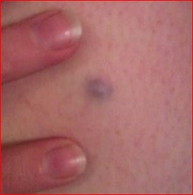
© Dannii Brighton, CC BY-SA 3.0, via Wikimedia Commons
Blue naevus symptoms (presentation)
Blue naevus usually arises during the second decade and do not change in shape or size thereafter.
Rarely, they can be present from birth.
If the cellular form of the lesion undergoes malignant transformation this usually manifests as a precipitate increase in size or, more rarely, as ulceration.6
Blue naevus can be found as pigmented lesions at unusual sites - eg, the female genitourinary tract,7 8 beneath nails, spermatic cord, bronchus, lymph nodes and prostate. Blue naevi found in the oral mucosa are rare but can have tendency to malignancy.9
Differential diagnosis for blue naevus10 11 12
Zurück zum InhaltMelanocytic naevus.
Combined naevus.
Compound naevus.
Neurofibroma.
Histiocytoma.
Tattooing effect (deliberate, or material accidentally pushed into the skin during trauma - eg, coalminer's tattoo, ink pens).
Thrombosed plantar wart.
Apocrine hydrocystoma.
Congenital naevus.
Granuloma telangiectaticum.
Naevi of Ota and Ito.
Lesen Sie unten weiter
Assoziierte Erkrankungen
Zurück zum InhaltCarney's syndrome/complex is a rare association of blue naevi with further abnormalities of the skin and other organs, inherited in an autosomal dominant fashion.
Blue naevus causes cardiac, endocrine, cutaneous and neural myxomatous tumours, plus a variety of pigmented lesions of the skin and mucosae.13
Untersuchungen
Zurück zum InhaltNone is usually required.
If the nature of a lesion is uncertain then dermoscopy may be performed by a dermatologist to distinguish it from melanomatous lesions.
Occasionally even dermoscopy is insufficient and biopsy may be required.14
Fluorescence in situ hybridisation (FISH) assay is sometimes needed to diagnose cellular blue naevi from blue naevus-like melanoma.15
Blue naevus treatment and management 16 17
Zurück zum InhaltTypical lesions with no other features that would suggest an alternative diagnosis, particularly melanoma, can be left alone, and the patient reassured.
However, as for any pigmented lesion, where there is doubt as to the diagnosis, it is safest to refer for dermatological advice.
There are occasional reports of recurrence of the lesion in a satellite form after excision; such lesions must be examined by further excision biopsy, preferably with dermatological opinion, to exclude malignant transformation.
Complications of blue naevus
Zurück zum InhaltCommon blue naevi do not have any complications, are benign and persist unchanged throughout life.
Cellular blue naevi are also usually benign but may, rarely, undergo malignant transformation.
Cellular naevi are larger and so more likely to present and undergo excision biopsy.
Prognose
Zurück zum InhaltThe prognosis for both types of lesion is excellent.
In the rare cases where cellular naevi become malignant then prognosis is improved by earlier diagnosis, as for melanoma.18
Exklusive Updates für medizinisches Fachpersonal
Bleiben Sie informiert mit den neuesten klinischen Updates, professionellen Einblicken und evidenzbasierten Leitlinien. Der Patient Pro-Newsletter stellt wesentliche Inhalte für Gesundheitsfachkräfte zusammen – direkt in Ihren Posteingang geliefert.
Durch das Abonnieren akzeptieren Sie unsere Datenschutzrichtlinie. Sie können sich jederzeit abmelden. Wir verkaufen Ihre Daten niemals.
Weiterführende Literatur und Referenzen
- Verbesserung der Ergebnisse für Menschen mit Hauttumoren, einschließlich Melanom; NICE-Leitlinien (Aktualisierung Mai 2010)
- Tièche-Jadassohn naevus; Whonamedit.com
- Sakamoto S, Oiso N, Narita T, et al; Blue nevus with a dermoscopic appearance of peripheral streaks with branches. Case Rep Dermatol. 2014 Feb 25;6(1):66-8. doi: 10.1159/000360215. eCollection 2014 Jan.
- Melanocytic naevus; DermNet NZ
- Jonjic N, Dekanic A, Glavan N, et al; Cellular Blue Nevus Diagnosed following Excision of Melanoma: A Challenge in Diagnosis. Case Rep Pathol. 2016;2016:8107671. doi: 10.1155/2016/8107671. Epub 2016 May 26.
- North JP, Yeh I, McCalmont TH, et al; Melanoma ex blue nevus: two cases resembling large plaque-type blue nevus with subcutaneous cellular nodules. J Cutan Pathol. 2012 Dec;39(12):1094-9. doi: 10.1111/cup.12015. Epub 2012 Nov 12.
- Leung AKC, Barankin B; An adolescent with a smooth, blue-black nodule on the dorsal wrist. Consultant Pediatricians. 2014;13(11):501-503.
- Lawrence F; Neonatal and Infant Dermatology, 2014.
- Kasturi S et al; Cellular blue nevus - A challenging entity. International Archives of Integrated Medicine, Vol. 2, Issue 2, February, 2015.
- Craddock KJ, Bandarchi B, Khalifa MA; Blue nevi of the Mullerian tract: case series and review of the literature. J Low Genit Tract Dis. 2007 Oct;11(4):284-9.
- Fitzhugh VA, Houck K, Heller DS; Vaginal blue nevus: report of a case and review of the literature. J Low Genit Tract Dis. 2011 Oct;15(4):325-7. doi: 10.1097/LGT.0b013e318213f3b8.
- Santos Tde S, Frota R, Martins-Filho PR, et al; Extensive intraoral blue nevus--case report. An Bras Dermatol. 2011 Jul-Aug;86(4 Suppl 1):S61-5.
- Blue Nevus; DermIS (Dermatology Information System), 2013
- Blue Nevus; American Osteopathic College of Dermatology
- Plensdorf S, Livieratos M, Dada N; Pigmentation Disorders: Diagnosis and Management. Am Fam Physician. 2017 Dec 15;96(12):797-804.
- Carney Complex, Type 1: CNC1; Online-Mendelsche-Erbfolge beim Menschen (OMIM)
- Di Cesare A, Sera F, Gulia A, et al; The spectrum of dermatoscopic patterns in blue nevi. J Am Acad Dermatol. 2012 Aug;67(2):199-205. doi: 10.1016/j.jaad.2011.08.018. Epub 2011 Oct 26.
- Gammon B, Beilfuss B, Guitart J, et al; Fluorescence in situ hybridization for distinguishing cellular blue nevi from blue nevus-like melanoma. J Cutan Pathol. 2011 Apr;38(4):335-41. doi: 10.1111/j.1600-0560.2010.01667.x. Epub 2011 Jan 19.
- Blue Naevus; Primary Care Dermatology Society, 2012
- Sardana K, Chakravarty P, Goel K; Optimal management of common acquired melanocytic nevi (moles): current perspectives. Clin Cosmet Investig Dermatol. 2014 Mar 19;7:89-103. doi: 10.2147/CCID.S57782. eCollection 2014.
- Damsky WE, Bosenberg M; Melanocytic nevi and melanoma: unraveling a complex relationship. Oncogene. 2017 Oct 19;36(42):5771-5792. doi: 10.1038/onc.2017.189. Epub 2017 Jun 12.
Lesen Sie unten weiter
Über den AutorVollständige Biografie anzeigen

Dr Colin Tidy, MRCGP
Allgemeinmediziner, Medizinischer Autor
MBBS, MRCGP, MRCP (Paediatrics), DCH
Dr. Colin Tidy ist ein NHS-Arzt mit Sitz in Oxfordshire.
Über den RezensentenVollständige Biografie anzeigen

Dr. Laurence Knott
Allgemeinmediziner, Medizinischer Autor
BSc (Hons) Biochemie, MBBS
Dr. Laurence Knott qualifizierte sich 1973 und hat umfangreiche Erfahrung als Allgemeinmediziner.
Artikelverlauf
Die Informationen auf dieser Seite wurden von qualifizierten Klinikern verfasst und begutachtet.
Next review due: 30 Mar 2027
31 Mar 2022 | Neueste Version

Fragen, teilen, verbinden.
Durchsuchen Sie Diskussionen, stellen Sie Fragen und teilen Sie Erfahrungen zu Hunderten von Gesundheitsthemen.

Fühlen Sie sich unwohl?
Bewerten Sie Ihre Symptome online kostenlos