Echokardiogramm
Begutachtet von Dr Colin Tidy, MRCGPZuletzt aktualisiert von Dr Toni Hazell, MRCGPZuletzt aktualisiert 22. Nov. 2022
Erfüllt die Anforderungen des Patienten Richtlinien des Patienten
- HerunterladenHerunterladen
- Teilen
- Language
- Diskussion
- Audio-Version
- Zu bevorzugten Quellen bei Google hinzufügen
An echocardiogram is an Ultraschall-Scan of the heart. It is sometimes just called an 'echo'. Ultrasound is a very high-frequency sound that you cannot hear but it can be emitted and detected by special machines. The scan can give accurate pictures of the heart muscle, the heart chambers and structures within the heart such as the valves.
Auf einen Blick
An echocardiogram uses ultrasound to create moving pictures of your heart.
Es kann zeigen, wie gut Ihr Herz und seine Klappen funktionieren.
The test is painless, takes 15-30 minutes, and requires no special preparation.
Eine Sonde wird mit Gel auf Ihrer Brust platziert, um Ultraschallwellen zu senden und zu empfangen.
Es gibt verschiedene Typen, einschließlich Doppler-, Stress- und transösophageale Echokardiogramme.
Results are sent to the doctor who requested the test.
What does an echocardiogram show?
An echocardiogram can be carried out for many different reasons. It may be done to check how well your heart is working after major heart problems such as a heart attack, or to look at how well the valves are moving inside the heart. An echocardiogram can also help to see any fluid that may have collected around the heart and may be used to see if symptoms such as shortness of breath are caused by a cardiac cause such as heart failure.
How accurate is a echocardiogram?
Interpreting an echocardiogram is a skilled job. The sonographer will interpret the images and write a report, which will be sent to the requesting clinician (GP or consultant). In some cases a consultant may want to carry out an echocardiogram themselves, to get a first-hand look at the images.
How is an echocardiogram done?
The test is painless and takes about 15-30 minutes. You may have to turn on to your side during the test so that the operator can scan the heart from different angles.
You will need to undress to the waist and lie on the couch. A probe is placed on your chest (it is a bit like a very thick blunt pen). Also, a substance called a contrast agent (lubricating jelly) is put on your chest so the probe makes good contact with the skin. The probe is connected by a wire to the ultrasound machine and monitor. Pulses of ultrasound are sent from the probe through the skin towards your heart. The ultrasound waves then 'bounce back' (echo) from the heart and various structures in the heart.
Echocardiogram procedure
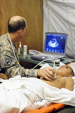
© Tech Sgt Luke Thelen, via Wikimedia Commons
The echoes are detected by the probe and are sent to the echocardiogram machine. They are displayed as a picture on the monitor. The picture is constantly updated so the scan can show movement as well as structure. (For example, the valves of a heart opening and closing.) The operator moves the probe around over the skin surface to obtain views from different angles. Some abnormalities can be seen quite clearly. For example, damaged heart valves, thickened heart muscle, some congenital heart defects, etc.
You do not need any special preparation before the test. You eat and drink normally before and after the test. Continue to take your usual medication.
Doppler echocardiography
This type of echocardiogram can measure variations in blood flow through your heart. For example, if can detect any abnormal blood flows next to a damaged valve. It can assess how well the heart valves are working. You do not need any special preparation before this test.
Stress echocardiogram
This test is done to show how well your heart responds to 'stress' such as exercise. In this test your doctor may do an echocardiogram, as described above, during or soon after exercise. Or you may be given a medication that causes the heart to beat harder and faster.
Transoesophageal echocardiogram
In this test you swallow a probe that is attached to a thin tube connecting it to an ultrasound machine. This views the heart from within the gullet (oesophagus) which lies just behind the heart. This can give a clearer view of the heart than normal echocardiography. It is done in situations where a very detailed picture is needed. For example, to assess valves before surgery is done to repair damaged valves, or to assess the extent of infection of a heart valve.
How long does it take to get echocardiogram results?
This varies locally. Results will go back to the requesting clinician and it is their responsibility to give them to the patient. So, if your echocardiogram was requested by a consultant, do not ring your GP for the result. Wait for your next consultant appointment, where you will get the result, or ring your consultant's secretary if you have a query. Hospital consultants should not ask patients to go to their GP for the results of tests that the consultant has requested.
Patientenauswahl für Herzuntersuchungen
Tests und Untersuchungen
Elektrokardiogramm
Ein Elektrokardiogramm (EKG) zeichnet die elektrische Aktivität des Herzens auf. Das Herz erzeugt winzige elektrische Impulse, die sich durch den Herzmuskel ausbreiten und das Herz zum Pumpen bringen. Diese Impulse können vom EKG-Gerät erkannt werden. Ein EKG kann dabei helfen, die Ursache von Symptomen wie dem Gefühl eines 'schlagenden Herzens' (Palpitationen) oder Brustschmerzen zu finden. Manchmal wird es im Rahmen routinemäßiger Untersuchungen durchgeführt – zum Beispiel bei Bluthochdruck oder vor einer Operation. Der EKG-Test ist schmerzfrei und harmlos. (Das EKG-Gerät zeichnet elektrische Impulse auf, die vom Körper kommen – es gibt keine Elektrizität in den Körper.)
von Dr. Colin Tidy, MRCGP

Tests und Untersuchungen
Koronarangiographie
Koronarangiographie ist ein spezieller Röntgentest, um detaillierte Informationen über Ihre Herzkranzgefäße zu erhalten. Er wird hauptsächlich bei Angina oder nach einem Herzinfarkt durchgeführt, um zu beurteilen, ob und welche Arterien blockiert sind und wie stark die Blockaden sind. Dabei handelt es sich um ein Verfahren namens Katheterisierung.
von Dr. Doug McKechnie, MRCGP
Häufig gestellte Fragen
Was ist der Unterschied zwischen einem Standard-Echokardiogramm und einem Doppler-Echokardiogramm?
Ein Standard-Echokardiogramm zeigt in erster Linie die Struktur und Bewegung Ihres Herzens. Ein Doppler-Echokardiogramm hingegen misst speziell die Variationen des Blutflusses durch Ihr Herz. Dadurch können abnormale Blutflüsse, beispielsweise neben einer beschädigten Klappe, erkannt und die Effektivität der Herzklappen beurteilt werden.
Wann wird ein transösophageales Echokardiogramm anstelle eines Standard-Echokardiogramms verwendet?
Ein transösophageales Echokardiogramm wird verwendet, wenn ein sehr detailliertes Bild des Herzens erforderlich ist. Dies liegt daran, dass die Sonde geschluckt wird und das Herz von innen aus der Speiseröhre betrachtet, die sich direkt hinter dem Herzen befindet, was eine klarere Sicht bietet als ein Standard-Echokardiogramm, bei dem die Sonde auf der Brust liegt. Es kann verwendet werden, um die Herzklappen vor einer Operation zu beurteilen oder das Ausmaß einer Herzklappeninfektion zu überprüfen.
Was passiert nach dem Echokardiogramm-Test?
Nach dem Test interpretiert der Sonograph die Bilder und erstellt einen Bericht. Dieser Bericht wird dann an den Kliniker gesendet, der das Echokardiogramm angefordert hat, wie z.B. Ihren Hausarzt oder einen Facharzt. Es liegt in deren Verantwortung, Ihnen die Ergebnisse mitzuteilen.
Muss ich etwas Besonderes tun, um mich auf ein Echokardiogramm vorzubereiten?
Nein, Sie benötigen keine spezielle Vorbereitung für ein Standard-, Doppler- oder Belastungs-Echokardiogramm. Sie können sowohl vor als auch nach dem Test normal essen und trinken und sollten Ihre regulären Medikamente wie gewohnt einnehmen.
Wie lange dauert ein Echokardiogramm normalerweise?
Der Test selbst ist in der Regel schnell und dauert normalerweise etwa 15 bis 30 Minuten.
Weiterführende Literatur und Referenzen
- Sengupta PP, Khandheria BK; Transoesophageal echocardiography. Heart. 2005 Apr;91(4):541-7.
- Mohamed AA, Arifi AA, Omran A; The basics of echocardiography. J Saudi Heart Assoc. 2010 Apr;22(2):71-6. doi: 10.1016/j.jsha.2010.02.011. Epub 2010 Mar 1.
Über den AutorVollständige Biografie anzeigen

Dr Toni Hazell, MRCGP
MBBS, BSc, MRCGP, DFSRH, Dip GU med, DRCOG, DCH (London, UK, 2000)
Dr. Toni Hazell hat ihren Abschluss an der St. Mary’s Hospital Medical School gemacht und ihr VTS am Northwick Park Hospital absolviert.
Über den RezensentenVollständige Biografie anzeigen

Dr Colin Tidy, MRCGP
Allgemeinmediziner, Medizinischer Autor
MBBS, MRCGP, MRCP (Paediatrics), DCH
Dr. Colin Tidy ist ein NHS-Arzt mit Sitz in Oxfordshire.
Artikelverlauf
Die Informationen auf dieser Seite wurden von qualifizierten Klinikern verfasst und begutachtet.
Artikel auch verfügbar in Englisch, Deutsch, Spanisch, Französisch, Italienisch, Portugiesisch, Hindi, Hebräisch, Arabisch, und Schwedisch.
Nächste Überprüfung fällig: 21. Nov. 2027
22. Nov. 2022 | Neueste Version

Fragen, teilen, verbinden.
Durchsuchen Sie Diskussionen, stellen Sie Fragen und teilen Sie Erfahrungen zu Hunderten von Gesundheitsthemen.

Fühlen Sie sich unwohl?
Bewerten Sie Ihre Symptome online kostenlos
Abonnieren Sie den Patienten-Newsletter
Ihre wöchentliche Dosis klarer, vertrauenswürdiger Gesundheitsberatung - geschrieben, um Ihnen zu helfen, sich informiert, selbstbewusst und in Kontrolle zu fühlen.
Durch das Abonnieren akzeptieren Sie unsere Datenschutzrichtlinie. Sie können sich jederzeit abmelden. Wir verkaufen Ihre Daten niemals.
Mehr zu Tests und Untersuchungen
- Arterielle Blutgase
- Audiologie
- Blutgerinnungstests
- Bluttests zur Erkennung von Entzündungen
- Darmkrebsvorsorge
- BRCA-Gene
- Koloskopie
- CT-Kolonographie
- DMSA-Scan
- Belastungstest
- Gallium-Scan
- Urinprobe aus dem Mittelstrahl
- Myokardperfusionsszintigraphie
- Proteinurie
- Radionuklid-Scan
- Sichelzellenmerkmal
- Sigmoidoskopie
- Symptomprüfer
- Thalassämie
- Ultraschalluntersuchung