DEXA-Scan
Begutachtet von Dr Hayley Willacy, FRCGP Zuletzt aktualisiert von Dr Colin Tidy, MRCGPZuletzt aktualisiert 20. März 2023
Erfüllt die Anforderungen des Patienten Richtlinien des Patienten
- HerunterladenHerunterladen
- Teilen
- Language
- Diskussion
- Audio-Version
- Zu bevorzugten Quellen bei Google hinzufügen
In dieser Serie:OsteoporoseBisphosphonateKalziumreiche LebensmittelVitamin-D-MangelVorbeugung von steroidinduzierter Osteoporose
DEXA scans (also called DXA scans or bone density scans) are used to check the density of bones. This test uses X-rays to show how strong bones are. A DEXA scan is different from a Knochenszintigrafie, which used radioactive chemicals to create a picture of the bones.
Auf einen Blick
A DEXA scan measures your bone density using low-energy X-rays.
Dense bones are stronger and less likely to break.
The scan usually takes 5 to 20 minutes while you lie on a couch.
DEXA scans use a very low level of X-ray radiation.
You do not need special preparation; avoid clothes with metal.
It may be advised if you have had a fracture or are at risk of 'thinning' bones.
DEXA scans are not advised for women who are pregnant.
Hinweis: Die folgenden Informationen dienen nur als allgemeine Richtlinie. Die Abläufe und die Durchführung von Tests können in verschiedenen Krankenhäusern variieren. Befolgen Sie stets die Anweisungen Ihres Arztes oder Ihres örtlichen Krankenhauses.
What is a DEXA scan?
DEXA stands for 'dual-energy X-ray absorptiometry'. DEXA (also sometimes known as DXA) is a test that measures the density of bones. Density means how much of something there is in a certain amount of space. The denser the tissue, the less X-rays pass through.
Air and water are less dense than solid things such as bone. This is because the particles which make air and water are not held closely together. In general, the more dense the bone, the stronger it is, and the less likely it is to break (fracture).
There are two different types of DEXA scanning devices:
Central DEXA devices are large machines that can measure bone density in the centre of your skeleton, such as your hip and spine.
Peripheral DEXA devices are smaller, portable machines that are used to measure bone density on the periphery of your skeleton, such as your wrist, heel or finger. These are mainly to get an idea about whether further tests are needed, as they are not as accurate as the larger DEXA machines.
How does a DEXA scan work?
A DEXA scan uses low-energy X-rays. A machine sends X-rays from two different sources through the bone being tested. Bone blocks a certain amount of the X-rays. The more dense the bone is, the less X-rays get through to the detector. By using two different X-ray sources rather than one it greatly improves the accuracy in measuring the bone density.
The amount of X-rays that comes through the bone from each of the two X-ray sources is measured by a detector. This information is sent to a computer which calculates a score of the average density of the bone. A low score indicates that the bone is less dense than it should be, some material of the bone has been lost and it is more prone to fracture.
How is a DEXA scan done?
You lie on your back on a couch and are asked to keep still while an X-ray detector (the 'scanner') comes over the area to be tested. An X-ray machine fires X-rays towards the detector. The bones commonly scanned are the bones of the back (the vertebrae), hip and wrist.
Smaller peripheral scanners are available in some places and can be used to check the bone mass density of the heel, wrist or finger.
How long does a DEXA scan take?
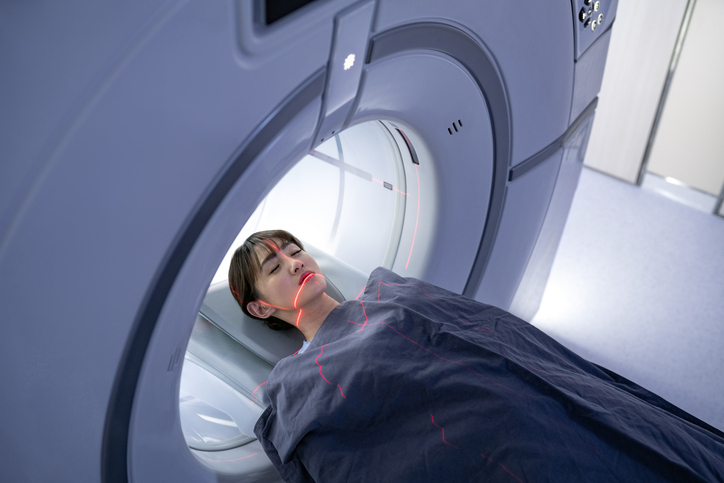
The scan usually takes between 5 and 20 minutes, depending on which part of your body is being examined and whether a central or peripheral scanner is being used. There is no 'tunnel' to pass through as there is in other types of scans such as an MRI or CT scan, so it should not affect people who do not like being in enclosed spaces.
DXA scanner
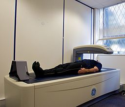
© Nick Smith photography (ALSPAC website), via Wikimedia Commons
Are DEXA scans safe?
DEXA scans use a very low level of X-ray radiation. This means it is safe for the technician doing the scan to stay in the room with you. (In standard X-ray tests, the technician has to stay behind a protective screen.)
Preparing for a DEXA scan
You do not need to do any special preparation prior to a DEXA scan. You can normally remain fully clothed, although you will need to avoid or remove clothes with metal in them (for example, zips, belts, buttons). You may also be asked to remove jewellery for the scan. In some places, you may be given a gown to wear.
Who should have a DEXA scan?
A DEXA scan may be advised if you have had a fracture of a bone after a minor injury. It may also be advised if you are considered at increased risk of 'thinning' of the bones (osteoporosis) and therefore at increased risk of having a fracture in future.
If your doctor thinks you have risk factors for osteoporosis, they may use a risk calculator such as one called FRAX® or QFracture®. This gives an idea of how likely you are to fracture your bones after a minor knock. If your risk is at a medium level, your doctor would then arrange a DEXA scan. This enables them to gain a clearer picture of your risk and then to decide whether you need any treatment.
DEXA scans are also used to monitor whether treatment for osteoporosis is working.
DEXA scans are not advised for women who are pregnant. You should also not have a DEXA scan within two weeks of certain other types of scans - for example, those using contrast dye.
For further information, see the separate leaflet called Osteoporose.
Patientenauswahl für Bildgebung

Tests und Untersuchungen
PET-Scan
Eine PET-Untersuchung erstellt Bilder, die zeigen, wo Zellen im Körper besonders aktiv sind. Sie wird am häufigsten zur Diagnose und Beurteilung von Krebs verwendet. Hinweis: Die nachstehenden Informationen sind nur eine allgemeine Orientierung. Die Abläufe und die Durchführung der Tests können in verschiedenen Krankenhäusern variieren. Befolgen Sie stets die Anweisungen Ihres Arztes oder des örtlichen Krankenhauses.
von Dr. Rosalyn Adleman, MRCGP
Tests und Untersuchungen
MRT-Scan
Eine MRT-Untersuchung ist ein sicherer und schmerzfreier Test, der detaillierte Bilder von Organen und anderen Strukturen in Ihrem Körper liefern kann. Hinweis: Die nachstehenden Informationen sind nur eine allgemeine Orientierung. Die Abläufe (und die Art der Durchführung der Tests) können zwischen verschiedenen Krankenhäusern variieren. Folgen Sie stets den Anweisungen Ihres Arztes oder des örtlichen Krankenhauses. Diese sind in der Regel mit Ihrem Terminbrief enthalten.
von Dr. Toni Hazell, MRCGP
Häufig gestellte Fragen
What is the purpose of a DEXA scan?
A DEXA scan is used to measure the density of bones. This measurement helps determine how strong your bones are and how likely they are to break (fracture). It can be advised if you've already had a fracture from a minor injury, or if you're considered at high risk of osteoporosis (thinning of the bones).
Does a bone density test show cancer?
The article states that a DEXA scan measures bone density to assess the risk of fractures or monitor osteoporosis treatment. It does not mention that a DEXA scan is used to detect cancer.
Do I need to do anything special to prepare for a DEXA scan?
You typically don't need any special preparation. You can usually remain fully clothed, but you will need to avoid or remove any clothing with metal, such as zips, belts, or buttons. You might also be asked to take off jewellery, and in some cases, you may be given a gown to wear.
Who should not have a DEXA scan?
DEXA scans are not recommended for pregnant women. Additionally, you should not have a DEXA scan within two weeks of having other types of scans that involve the use of contrast dye.
What is the difference between central and peripheral DEXA devices?
Central DEXA devices are large machines that measure bone density in the core parts of your skeleton, like your hip and spine. Peripheral DEXA devices are smaller, portable machines used for areas like the wrist, heel, or finger. Peripheral devices mainly indicate if further testing is needed, as they are not as accurate as the larger central machines.
Weiterführende Literatur und Referenzen
- Osteoporose: Einschätzung des Risikos für Knochenbrüche bei Schwäche; NICE-Klinikleitlinie (August 2012, aktualisiert Februar 2017)
- Behandlung von Osteoporose und Vorbeugung von Fragilitätsfrakturen – Eine nationale klinische Leitlinie; Scottish Intercollegiate Guidelines Network (SIGN - Januar 2021)
- Osteoporose - Vorbeugung von Fragilitätsfrakturen; NICE CKS, Juli 2021 (nur für UK-Zugang)
- Klinische Leitlinie zur Prävention und Behandlung von Osteoporose; Nationale Leitlinie Osteoporose (aktualisiert September 2021)
Über den AutorVollständige Biografie anzeigen

Dr Colin Tidy, MRCGP
Allgemeinmediziner, Medizinischer Autor
MBBS, MRCGP, MRCP (Paediatrics), DCH
Dr. Colin Tidy ist ein NHS-Arzt mit Sitz in Oxfordshire.
Über den RezensentenVollständige Biografie anzeigen

Dr Hayley Willacy, FRCGP
Allgemeinmediziner, Medizinischer Autor
MBChB (1992), DRCOG, DFFP, MRCOG (Part 1) MRCGP (2007), DFSRH (2013), MSc - medical education (2020)
Dr. Hayley Willacy war eine NHS-Hausärztin, die in Nordwestengland arbeitete und 2022 nach 30 Jahren aus der klinischen Praxis ausschied.
Artikelverlauf
Die Informationen auf dieser Seite wurden von qualifizierten Klinikern verfasst und begutachtet.
Artikel auch verfügbar in Englisch, Deutsch, Spanisch, Französisch, Italienisch, Portugiesisch, Hindi, Hebräisch, Arabisch, und Schwedisch.
Nächste Überprüfung fällig: 18. März 2028
20. März 2023 | Neueste Version

Fragen, teilen, verbinden.
Durchsuchen Sie Diskussionen, stellen Sie Fragen und teilen Sie Erfahrungen zu Hunderten von Gesundheitsthemen.

Fühlen Sie sich unwohl?
Bewerten Sie Ihre Symptome online kostenlos
Abonnieren Sie den Patienten-Newsletter
Ihre wöchentliche Dosis klarer, vertrauenswürdiger Gesundheitsberatung - geschrieben, um Ihnen zu helfen, sich informiert, selbstbewusst und in Kontrolle zu fühlen.
Durch das Abonnieren akzeptieren Sie unsere Datenschutzrichtlinie. Sie können sich jederzeit abmelden. Wir verkaufen Ihre Daten niemals.
Mehr zu Tests und Untersuchungen
- Audiologie
- Blutgerinnungstests
- Blutgruppen und -typen
- Bluttests
- Kardiale Enzyme
- Echokardiogramm
- Endoskopischer Ultraschall-Scan
- Vollblutbild und Blutausstrich
- Heim- und ambulante Blutdruckaufzeichnung
- Hysterosalpingographie
- Intravenöse Urographie
- Nierenbiopsie
- Leberbiopsie
- Lumbalpunktion
- Myokardperfusionsszintigraphie
- Neugeborenen-Bluttest
- Neugeborenen-Untersuchungen
- Sichelzellenmerkmal
- Symptomprüfer
- Synacthen-Test