MRT-Scan
Begutachtet von Dr Colin Tidy, MRCGPZuletzt aktualisiert von Dr Toni Hazell, FRCGPZuletzt aktualisiert 7. Aug. 2023
Erfüllt die Anforderungen des Patienten Richtlinien des Patienten
- HerunterladenHerunterladen
- Teilen
- Language
- Diskussion
- Audio-Version
- Zu bevorzugten Quellen bei Google hinzufügen
Eine MRT-Untersuchung ist eine sichere und schmerzfreie Untersuchung, die detaillierte Bilder von Organen und anderen Strukturen in Ihrem Körper liefern kann.
Hinweis: Die nachstehenden Informationen sind nur eine allgemeine Orientierung. Die Abläufe (und die Art der Durchführung der Tests) können zwischen verschiedenen Krankenhäusern variieren. Folgen Sie stets den Anweisungen Ihres Arztes oder des örtlichen Krankenhauses. Diese sind in der Regel mit Ihrem Terminbrief enthalten.
Auf einen Blick
An MRI scan uses a strong magnetic field and radio waves to create detailed images inside your body.
It can show different tissues, organs, and other structures, aiding in diagnosing various conditions.
You lie on a couch inside a tunnel-like scanner, and you must keep still during the 15-40 minute scan.
The scan is painless but can be noisy, so you will be offered headphones or earplugs.
Inform your doctor if you have fear of enclosed spaces or any metal implants due to the strong magnet.
Pregnant women are usually advised against an MRI unless urgent, as long-term effects on a baby are unknown.
Was ist eine MRT-Untersuchung?
Die MRT steht für Magnetresonanztomographie. Eine MRT-Untersuchung verwendet ein starkes Magnetfeld und Radiowellen, um ein detailliertes Bild auf einem Computerbildschirm zu erstellen. Sie kann verschiedene Gewebearten, Organe und andere Strukturen in Ihrem Körper sichtbar machen.
Dieses Bild ist eine MRT-Aufnahme eines Gehirns. Die Augäpfel der Person sind am oberen Rand des Bildes sichtbar.
MRT-Scan
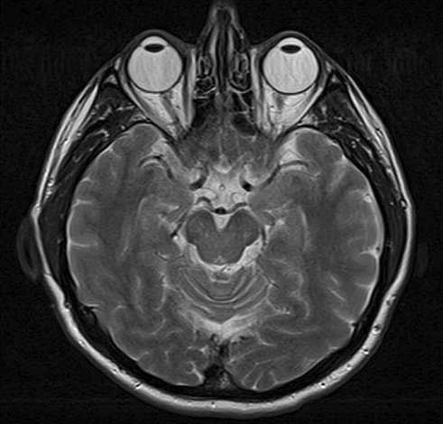
© Von Ptrump (Eigenarbeit) über Wikimedia Commons
Wie funktioniert ein MRT-Scan?
Ihr Körper enthält Millionen von Wasserstoffatomen. Wenn Sie in einem MRT-Scanner sind:
Ein starkes Magnetfeld richtet Teilchen namens Protonen aus, die sich in den Wasserstoffatomen befinden. Alle Protonen richten sich parallel zum Magnetfeld aus, wie winzige Magnete. (Normalerweise liegen die Millionen von Protonen in zufälligen Richtungen.)
Dann werden kurze Impulse von Radiowellen vom Scanner in Ihren Körper gesendet. Die Radiowellen stoßen die Protonen aus ihrer Position.
Wenn der Ausbruch der Radiowellen stoppt, richten sich die Protonen wieder aus. Dabei senden sie Radiowellen aus. Die Protonen in verschiedenen Geweben des Körpers richten sich mit unterschiedlichen Geschwindigkeiten wieder aus. Daher variiert das Signal, das von verschiedenen Körpergeweben ausgesendet wird. Zum Beispiel können weichere Gewebe anhand der ausgesendeten Signale von härteren Geweben unterschieden werden.
Diese Signale werden von einem Empfangsgerät im Scanner erfasst.
Das Empfangsgerät überträgt die Signale an einen Computer. Der Computer erstellt ein Bild basierend auf den vom Körper ausgesendeten Funksignalen.
Was beinhaltet eine MRT-Untersuchung?
Der MRT-Scanner ist wie ein etwa 1,5 Meter langer Tunnel, umgeben von einem großen kreisförmigen Magneten. Sie liegen auf einer Liege, die dann in den Scanner geschoben wird. Ein 'Empfangsgerät', ähnlich einer Antenne, wird hinter oder um den zu untersuchenden Körperteil platziert. Dieses erkennt die winzigen Radiosignale, die von Ihrem Körper ausgesendet werden. Wenn ein Bild aufgenommen wird, müssen Sie für einige Minuten stillhalten, sonst könnte das Scan-Bild verschwommen sein.
MRT-Scanner
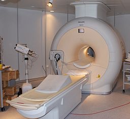
© Von Jan Ainali (eigene Arbeit) über Wikimedia Commons.
Die Untersuchung selbst ist schmerzfrei .Der gesamte Vorgang kann 15-40 Minuten dauern. Es kann etwas unangenehm sein, während dieser Zeit ruhig auf der Couch zu liegen. Kleine Kinder benötigen möglicherweise eine Vollnarkose, um sie lange genug ruhig zu halten, damit die Bilder gemacht werden können. Der Ort, an dem Sie liegen, ist ziemlich eingezäunt, und manche Menschen könnten das sehr beunruhigend finden.
Wenn Sie Angst vor beengten Räumen haben (Klaustrophobie) Sie sollten dies vor der Untersuchung mit Ihrem Arzt besprechen. In einigen Regionen des Landes gibt es Zugang zu 'offenen' Scannern. Diese sind jedoch nicht weit verbreitet, und die Wartezeiten für einen 'offenen' Scanner sind in der Regel länger als bei einem geschlossenen Scanner.
In einigen Fällen wird eine Injektion eines speziellen Kontrastmittels in den Blutkreislauf über eine Vene im Arm verabreicht. Dies hilft, klarere Bilder bestimmter Gewebe oder Organe zu erhalten, die untersucht werden.
Der Radiologietechnologe sitzt im Kontrollraum neben dem Scanner und beobachtet durch das Fenster. Sie können jedoch mit ihm sprechen, meist über eine Gegensprechanlage, und Sie werden jederzeit auf einem Monitor überwacht.
Der Scanner ist laut, daher erhalten Sie Kopfhörer oder Ohrstöpsel, um Ihre Ohren vor dem Lärm zu schützen. Häufig können Sie über die Kopfhörer Radio oder Musik hören.
Wofür wird eine MRT-Untersuchung verwendet?
An MRI scan can create clear pictures of most parts of the body. So, it is useful for all sorts of reasons where other tests (such as Röntgenaufnahmen) geben nicht die erforderlichen Informationen an.
Es wird häufig verwendet, um detaillierte Bilder des Gehirns und des Rückenmarks zu erhalten, Anomalien und Tumore zu erkennen. Selbst gerissene Bänder um die Gelenke können mit einer MRT-Untersuchung entdeckt werden. Daher wird es nach Sportverletzungen immer häufiger eingesetzt.
Gehirn
MRI is the first-choice investigation for Gehirntumoren, as it produces clearer images than computerised tomography (CT) and shows hard-to-reach areas of the brain. There is clear contrast between grey and white matter parts of the brain and this makes MRI the best choice for many other conditions, including Multiple Sklerose, Schlaganfall, Alzheimer-Krankheit und Epilepsie.
Bewegungsapparat
Hier wird die MRT verwendet, um die Wirbelsäule zu untersuchen – zur Beurteilung von Gelenkerkrankungen, Nervendruck und Weichteiltumoren.
Gastrointestinal system
MRI allows non-invasive assessment of entzündliche Darmerkrankung und Darmtumore. Es kann auch Probleme in der Leber und Bauchspeicheldrüse erkennen.
Blutgefäße und das Herz
Dies wird als Magnetresonanzangiographie (MRA) bezeichnet und erstellt Bilder der Arterien, um abnormale Verengungen oder Gefäßwanddilatationen (die Gefahr des Platzen haben) zu erkennen. Die MRA wird häufig zur Beurteilung der Arterien im Hals und Gehirn, der thorakalen und abdominalen Aorta, der Nierenarterien und der Beine verwendet. Sie kann auch zur Beurteilung angeborener Herzfehler eingesetzt werden.
Vorbereitung auf eine MRT-Untersuchung
Ihr örtliches Krankenhaus sollte Ihnen Informationen darüber geben, was vor der Untersuchung erforderlich ist.
Der MRT-Scanner verwendet einen äußerst starken Magneten, daher können Personen mit bestimmten Arten von medizinischen Implantaten nicht untersucht werden. Dies liegt daran, dass der Magnet medizinische Geräte mit Metall darin möglicherweise verschieben oder deren Funktion beeinträchtigen kann.
Daher sollten Sie vor dem Betreten des Scanning-Bereichs gefragt werden, ob Sie medizinische Geräte in Ihrem Körper haben. Möglicherweise müssen Sie einen Sicherheitsfragebogen ausfüllen, der nach Gegenständen fragt, die Metall enthalten könnten. Die folgende Liste ist keine endgültige Aufzählung, sondern soll Ihnen helfen, sich an die Arten von Dingen zu erinnern, die Radiografen wissen müssen:
Internes (implantiertes) Defibrillator oder Herzschrittmacher.
Ohr ( Cochlea )-Implantat.
Chirurgische Clips wie die, die bei Gehirnaneurysmen verwendet werden.
Künstliche Herzklappen.
Implantierte Medikamenten-Infusionsports.
Künstliche Gliedmaßen oder metallische Gelenke.
Implantierte Nervenstimulatoren.
Stifte, Schrauben, Platten, Stents oder chirurgische Klammern.
Es ist auch wichtig, dem Radiologen mitzuteilen, wenn Sie jemals Metallteile in Ihren Augen oder Ihrem Körper gehabt haben. In einigen Fällen benötigen Sie möglicherweise vor einer MRT-Untersuchung eine Röntgenaufnahme, um sicherzustellen, dass Sie sicher in den Scanner gelangen können.
Nebenwirkungen einer MRT-Untersuchung
MRI scans are painless and thought to be safe. MRI scans do not use X-rays so the possible concerns associated with X-ray pictures and CT-Scans (which use X-rays) are not associated with MRI scans.
Allerdings:
Selten reagieren manche Menschen auf das manchmal verwendete Kontrastmittel.
Schwangeren Frauen wird normalerweise geraten, sich keiner MRT-Untersuchung zu unterziehen, es sei denn, es ist dringend erforderlich. Obwohl die Untersuchung als sicher gilt, sind die langfristigen Auswirkungen starker Magnetfelder auf ein sich entwickelndes Baby noch nicht bekannt.
Was Sie nach der MRT-Untersuchung erwarten können
Es gibt keine Nachwirkungen der Untersuchung. Sie können Ihre normalen Aktivitäten sofort nach Abschluss der Untersuchung wieder aufnehmen. Die Bilder der Untersuchung werden von einem Spezialisten, einem Radiologen, ausgewertet, der einen Bericht an den Arzt sendet, der die Untersuchung angeordnet hat.
Es ist üblich, mindestens zwei Wochen auf Ihre Ergebnisse zu warten. Falls es dringende Befunde gibt, wird der Facharzt so schnell wie möglich informiert. Das Ergebnis geht an die Person zurück, die die Untersuchung angefordert hat. Wenn ein Krankenhausarzt die Untersuchung angefordert hat, erhalten Sie die Ergebnisse bei Ihrem nächsten Termin bei ihm. Wenn Sie besorgt sind, können Sie die Sekretärin anrufen, aber wenden Sie sich nicht an den Hausarzt, um das Ergebnis zu erfragen, da dieser es nicht haben wird. Wenn der Hausarzt eine MRT angefordert hat, erhalten Sie die Ergebnisse von ihm.
Patientenauswahl für Bildgebung

Tests und Untersuchungen
Röntgenuntersuchung
Röntgenuntersuchungen zeigen Knochen und bestimmte andere Gewebe.
von Dr. Hayley Willacy, FRCGP

Tests und Untersuchungen
PET-Scan
Eine PET-Untersuchung erstellt Bilder, die zeigen, wo Zellen im Körper besonders aktiv sind. Sie wird am häufigsten zur Diagnose und Beurteilung von Krebs verwendet. Hinweis: Die nachstehenden Informationen sind nur eine allgemeine Orientierung. Die Abläufe und die Durchführung der Tests können in verschiedenen Krankenhäusern variieren. Befolgen Sie stets die Anweisungen Ihres Arztes oder des örtlichen Krankenhauses.
von Dr. Rosalyn Adleman, MRCGP
Häufig gestellte Fragen
Can I drive a car immediately after having an MRI scan?
Yes, there are no after-effects from an MRI scan, so you can usually return to your normal activities, including driving, as soon as the scan is finished.
How long will it take to get my MRI scan results?
It usually takes at least two weeks to receive your MRI scan results. However, if there are urgent findings, the specialist will inform the requesting doctor as soon as possible. The results will be sent to the doctor who requested the scan; if it was a hospital consultant, you'll get them at your next appointment with them. If your GP requested the MRI, they will provide you with the results.
What should I do if I have a fear of enclosed spaces?
If you have a fear of confined spaces (claustrophobia), you should discuss this with your doctor before your scan. While 'open' scan machines exist, they are not widely available and usually involve a longer waiting period compared to the more common enclosed scanners.
Why would I be given an injection during an MRI scan?
In some cases, a special contrast dye is injected into your bloodstream through a vein in your arm. This dye helps to produce clearer pictures of specific tissues or organs that are being examined during the scan.
Will I be alone during the MRI scan?
No, you will not be alone. The radiographer will be in a control room next to the scanner, observing you through a window and on a monitor at all times. You can communicate with them, usually via an intercom system.
Weiterführende Literatur und Referenzen
- MRT-Scan; Krebsforschung UK; 2022
Über den AutorVollständige Biografie anzeigen

Dr Toni Hazell, FRCGP
MBBS, BSc, FRCGP, DFSRH, Dip GU med, DRCOG, DCH (London, UK, 2000)
Dr. Toni Hazell hat ihren Abschluss an der St. Mary’s Hospital Medical School gemacht und ihr VTS am Northwick Park Hospital absolviert.
Über den RezensentenVollständige Biografie anzeigen

Dr Colin Tidy, MRCGP
Allgemeinmediziner, Medizinischer Autor
MBBS, MRCGP, MRCP (Paediatrics), DCH
Dr. Colin Tidy ist ein NHS-Arzt mit Sitz in Oxfordshire.
Artikelverlauf
Die Informationen auf dieser Seite wurden von qualifizierten Klinikern verfasst und begutachtet.
Artikel auch verfügbar in Englisch, Deutsch, Spanisch, Französisch, Italienisch, Portugiesisch, Hindi, Hebräisch, Arabisch, und Schwedisch.
Nächste Überprüfung fällig: 5. Aug. 2028
7. Aug. 2023 | Neueste Version

Fragen, teilen, verbinden.
Durchsuchen Sie Diskussionen, stellen Sie Fragen und teilen Sie Erfahrungen zu Hunderten von Gesundheitsthemen.

Fühlen Sie sich unwohl?
Bewerten Sie Ihre Symptome online kostenlos
Abonnieren Sie den Patienten-Newsletter
Ihre wöchentliche Dosis klarer, vertrauenswürdiger Gesundheitsberatung - geschrieben, um Ihnen zu helfen, sich informiert, selbstbewusst und in Kontrolle zu fühlen.
Durch das Abonnieren akzeptieren Sie unsere Datenschutzrichtlinie. Sie können sich jederzeit abmelden. Wir verkaufen Ihre Daten niemals.
Mehr zu Tests und Untersuchungen
- Antikörper- und Antigentests
- Audiologie
- Biopsie
- Blutgerinnungstests
- Bluttests zur Erkennung von Entzündungen
- Knochenszintigraphie
- BRCA-Gene
- Zerebrale Angiographie
- CT-Scan
- Zystoskopie
- Elektroenzephalograph
- Endoskopischer Ultraschall-Scan
- Feinnadelaspiration
- Intravenöse Urographie
- MRCP-Scan
- Neugeborenen-Hörtest
- Proteinurie
- Routinemäßiger Bluttest zur Nierenfunktion
- Spirometrie
- Ultraschalluntersuchung