Shin splints
Medial tibial stress syndrome
Peer reviewed by Dr Colin Tidy, MRCGPLast updated by Dr Doug McKechnie, MRCGPLast updated 13 Mar 2023
Meets Patient’s editorial guidelines
- DownloadDownload
- Share
- Language
- Discussion
- Audio Version
- Add to preferred sources on Google
In this series:Compartment syndrome
The term 'shin splints' refers to pain pain in the lower leg, specifically along the front of the leg between the knee and the ankle - the area known as the shin. The exact cause of shin splints is not certain but they tend to be as a result of overuse and typically occur in runners. Rest is the most important treatment. Shock-absorbent insoles in your training shoes, graduated running programmes and regularly replacing your training footwear may help in prevention.
At a glance
Shin splints are pain in the lower leg, specifically along the front of the shin bone.
The main symptom is pain and tenderness in the middle and lower shin, often on the inner half.
Pain often starts after running or exercising, but can occur during activity as it worsens.
They are thought to be caused by overuse or overactivity, especially in runners.
Treatment involves rest, activity modification, ice, elevation, and painkillers.
Shin splints usually improve within two to four weeks of stopping the activity.
Shock-absorbent shoe inserts and replacing running shoes regularly may help prevent them.
What are shin splints?
Shin splints
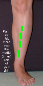
Shin splints are the name often given to exercise-induced pain in the lower leg, specifically along the front of the leg between the knee and the ankle - the area known as the shin bone.
What are the symptoms of shin splints?
Shin splints are really a symptom rather than a specific diagnosis because they are probably caused by a number of different problems. Shin splints are one of the most common problems in the lower leg in people who exercise or play sports. In typical shin splints, pain is felt more over the inner (medial) part of your shin.
The main symptom is pain and tenderness in the shin area. The pain tends to be in the middle and lower shin and on the inner (medial) half. Pain first comes on after running or exercising. However, over time, the pain can come on during running or exercising. If severe, it may also come on when climbing stairs.
Shin splints are sometimes called medial tibial stress syndrome.
What causes shin splints?
Experts do not all agree on the cause of shin splints and the exact cause is not known. They are thought to be caused by overuse or overactivity and typically occur in runners. There are certain things that have been suggested that may make shin splints more likely. These include:
A sudden increase in training frequency or intensity.
A lack of calcium.
Hard running surfaces.
Running up an incline
Previous leg injury.
Poorly fitted or inadequate running shoes that do not support the foot and ankle.
Various problems with leg muscles such as the lower leg and in foot position, including over-pronation of the foot. (The foot (and ankle) normally roll slightly inwards when we move. In over-pronation, the foot rolls inwards more than normal.)
Flat feet. These are a cause of over-pronation as well.
Some experts suggest that shin splints are caused by small tears in the structure of the membrane between the two bones of the leg below the knee (the tibia and fibula). This structure is called the interosseous membrane.
Others suggest that they may also be caused by:
Overuse injury of tendons (tendinopathy).
Muscle sprains.
Inflammation of the membrane surrounding the tibia and fibula bones (periostitis).
Tiny fractures (microfractures) in the surface of the tibia have also been suggested as a cause.
The relationship between shin splints and compartment syndrome is not clear. Some experts consider shin splints to be a form of compartment syndrome, whereas others believe that compartment syndrome is a separate condition that can cause shin splints. See separate leaflet called Compartment Syndrome for more details.
How to help shin splints
Rest and activity modification
This is the main treatment for shin splints. This means reducing, and often completely avoiding, any activity, such as running, that may have led to the shin splints. Switching to other exercises that don't involve repetitive, high-impact activity - such as cycling or swimming - can be a good way to maintain fitness.
Once recovered, you may be able to return to your normal activities, but should build up gradually and avoid over-training.
For athletes, physiotherapists might give advice on how to improve technique when jumping or running, to reduce the risk of shin splints coming back.
Ice
Applying ice to your shin and raising your leg may also help to relieve pain from shin splints. You can make a cold pack by wrapping ice cubes in a plastic bag or towel. (Do not put ice directly next to skin as it may cause an ice burn.) A bag of frozen peas is an alternative.
Gently press the ice pack on to the injured part. The cold from the ice is thought to reduce blood flow to the damaged ligament. This may limit pain and inflammation. After the first application, some doctors recommend re-applying for 10-20 minutes every two hours (during the daytime) for the first 48 hours.
Elevation
Elevation of the leg aims to limit and reduce any swelling. Keep the foot up on a chair to at least hip level when you are sitting. (It may be easier to lie on a sofa and to put your foot on some cushions.) When you are in bed, put your foot on a pillow.
Painkillers
Painkillers such as paracetamol are useful to ease pain. It is best to take these regularly initially. Anti-inflammatory painkillers are an alternative. There are many types and brands - for example, ibuprofen. They limit inflammation andmay also relieve pain and swelling.
Side-effects sometimes occur with anti-inflammatory painkillers. Stomach pain, and bleeding from the stomach are the most serious. Some people with asthma, high blood pressure, kidney failure and heart failure may not be able to take anti-inflammatory painkillers.
A plaster cast
This may be recommended in severe cases.
Surgery
This may be considered if all else fails. The operation is called a fasciotomy. It involves making an incision in the tissue overlying the muscles of the lower part of the leg (the posterior compartment).
It's very rare for shin splints to need surgery.
How long do shin splints take to heal?
Shin splints usually improve within two to four weeks of stopping the activity that was causing them. It can take longer to return to your previous level of exercise after that, especially if you're building up gradually. Some people take longer to recover.
How to avoid shin splints
Studies and trials have been done to look at preventing shin splints. No single prevention method has been found to be consistently effective and further trials are needed. However, one of the things that does show some promise is the use of shock-absorbent shoe inserts (insoles) inside shoes while you are exercising. Special insoles to correct over-pronation of your foot (if you have this) may also be helpful. You should also regularly replace your running shoes. Graduated running programmes that build in rest days may also help.
What else could the pain be?
Stress fractures of one of the two bones in the leg below the knee (the tibia) can also cause shin splint-type pain. A stress fracture is a type of incomplete fracture in a bone. Stress fractures tend to occur as a result of overuse and are known as overuse injuries.
How to test for shin splints
Shin splints can be diagnosed on the basis of typical symptoms and findings on examination. Tests aren't required, unless the diagnosis isn't clear, or there's thought to be a possibility of another problem, like a stress fracture.
X-rays of the affected area, taken as part of any investigative work-up in people with shin splints, are typically reported as being entirely normal. The main reason that your doctor may suggest an X-ray of your leg below the knee is to rule out a stress fracture in one of the bones. However, not all stress fractures show up on X-rays.
In some cases, your doctor may refer you for a bone scan of your lower leg. This can help to differentiate between shin splints and a stress fracture. A bone scan involves an injection of a very small amount of radioactive material, usually into a vein in your arm.
A gamma camera is then used that can detect the radiation emitted by the injected material. This can show up a stress fracture or changes that can occur in the bones in shin splints. A magnetic resonance imaging (MRI) scan is also sometimes used to help to tell the difference between shin splints and stress fractures.
There may be a link between low vitamin D and shin splints. A blood test to check vitamin D levels might be recommended if shin splints keep coming back, or are otherwise proving difficult to treat.
What is the outlook (prognosis)?
With rest and treatment, you can fully recover from shin splints. However, they may return (recur) if you do not look at the underlying cause of your shin splints.
When you have recovered from your shin splints, you may benefit from seeing a specialist such as a sports physiotherapist or a podiatrist. They may be able to help you to modify your exercise routine and may also be able to assess your legs, feet and shoes. This may show up some problems that may have caused your shin splints, such as over-pronation of your foot or poorly fitting training shoes. They may suggest the insertion of an insole inside your shoes.
Patient picks for Sports injuries

Bones, joints and muscles
Hamstring injury
A hamstring injury is a strain (tear) to one or more of the three large muscles at the back of the thigh (or their tendons at the back of the knee or in the pelvis).
by Dr Toni Hazell, FRCGP

Bones, joints and muscles
Groin strain
Anyone can experience having a pulled groin (groin strain) but it's much more common when running, jumping and playing sports.
by Dr Hayley Willacy, FRCGP
Frequently asked questions
Can I continue exercising if I have shin splints, or should I stop completely?
It is generally recommended to reduce, or even completely avoid, activities that caused your shin splints, particularly those involving repetitive, high-impact movements like running. You can switch to other exercises that don't put stress on your shins, such as cycling or swimming, to maintain your fitness during recovery.
What is the difference between shin splints and compartment syndrome?
The relationship between shin splints and compartment syndrome is not fully clear. Some experts consider shin splints to be a form of compartment syndrome, while others view compartment syndrome as a separate condition that can cause symptoms similar to shin splints.
How do specialists like physiotherapists or podiatrists help with shin splints?
After you've recovered, a sports physiotherapist can offer advice on improving your running or jumping technique to help prevent shin splints from returning. A podiatrist can assess your legs, feet, and shoes to identify issues like over-pronation or ill-fitting footwear that might have contributed to your shin splints, and suggest solutions like insoles.
What options are available if my shin splints are very severe and don't get better with rest?
In very severe cases, a plaster cast might be recommended. If all other treatments fail, surgery called a fasciotomy may be considered. This involves making an incision in the tissue overlying the muscles in the lower leg. However, surgery is rarely needed for shin splints.
If I get shin splints repeatedly, could there be an underlying reason?
If shin splints keep recurring or are difficult to treat, your doctor might recommend a blood test to check your vitamin D levels, as there could be a link between low vitamin D and persistent shin splints.
Further reading and references
- Shin Splints/Medial Tibial Stress Syndrome; Wheeless' Textbook of Orthopaedics
- Craig DI; Current developments concerning medial tibial stress syndrome. Phys Sportsmed. 2009 Dec;37(4):39-44.
- Winkelmann ZK, Anderson D, Games KE, et al; Risk Factors for Medial Tibial Stress Syndrome in Active Individuals: An Evidence-Based Review. J Athl Train. 2016 Dec;51(12):1049-1052. doi: 10.4085/1062-6050-51.12.13. Epub 2016 Nov 11.
- Moen MH, Holtslag L, Bakker E, et al; The treatment of medial tibial stress syndrome in athletes; a randomized clinical trial. Sports Med Arthrosc Rehabil Ther Technol. 2012 Mar 30;4:12. doi: 10.1186/1758-2555-4-12.
- Menendez C, Batalla L, Prieto A, et al; Medial Tibial Stress Syndrome in Novice and Recreational Runners: A Systematic Review. Int J Environ Res Public Health. 2020 Oct 13;17(20):7457. doi: 10.3390/ijerph17207457.
About the authorView full bio

Dr Doug McKechnie, MRCGP
Medical Writer
MA, MBBS, MSc, DRCOG, MRCP(UK), MRCGP(2021), FHEA
Dr Doug McKechnie is an NHS GP working in London. He works full-time clinically and is also the Deputy Lead for the Clinical and Professional Practice module at University College London Medical School.
About the reviewerView full bio

Dr Colin Tidy, MRCGP
General Practitioner, Medical Author
MBBS, MRCGP, MRCP (Paediatrics), DCH
Dr Colin Tidy is an NHS Doctor, based in Oxfordshire.
Article history
Article also available in English, German, Spanish, French, Italian, Portuguese, Hindi, Hebrew, Arabic, and Swedish.
Next review due: 11 Mar 2028
13 Mar 2023 | Latest version
Join the conversation
Share your thoughts or experiences with others in our patient communities.

Ask, share, connect.
Browse discussions, ask questions, and share experiences across hundreds of health topics.

Feeling unwell?
Assess your symptoms online for free
Sign up to the Patient newsletter
Your weekly dose of clear, trustworthy health advice - written to help you feel informed, confident and in control.
By subscribing you accept our Privacy Policy. You can unsubscribe at any time. We never sell your data.