Glaskörperblutung
Begutachtet von Dr Doug McKechnie, MRCGPZuletzt aktualisiert von Dr Colin Tidy, MRCGPLast updated 29 Jun 2024
Erfüllt die Anforderungen des Patienten Richtlinien des Patienten
- HerunterladenHerunterladen
- Teilen
- Language
- Diskussion
- Audio-Version
- Add to preferred sources on Google
In dieser Serie:SehproblemeMakuladegenerationAugenflimmern, Blitze und HalosNetzhautvenenverschlussRiesenzellarteriitisSchielen bei Kindern
Vitreous haemorrhage is bleeding into the jelly-like filling of the back part of your eye. This substance is the vitreous humour. It helps the eye keep its shape and is normally clear, allowing light from outside the eye to pass through it to reach the retina.
Vitreous haemorrhage varies in degree from mild, with 'floaters' and haziness in the vision, to complete loss of vision. It is painless and it comes on quite quickly. Usually only one eye is affected. Whilst it is very alarming, once the bleeding has been treated, many cases resolve and vision is restored to where it was before.
At a glance
Vitreous haemorrhage is when blood leaks into the jelly-like substance in your eye.
This can cause floaters, blurred vision, or total loss of sight.
Common causes include advanced diabetic eye disease, retinal tears, and eye trauma.
Diagnosis involves an eye examination, sometimes with an ultrasound or angiogram.
Treatment depends on the cause and may involve laser, injections, or surgery.
If you have sudden vision loss, see a GP, optician, or A&E immediately.
In diesem Artikel:
Video picks for Augenkrankheiten
Lesen Sie unten weiter
What is vitreous haemorrhage?
Vitreous haemorrhage occurs when blood leaks into the vitreous humour inside the eye. The leaked blood most commonly comes from blood vessels at the back of the eye (retinal blood vessels). This is more likely to happen if the blood vessels have been damaged (eg, by trauma) or are particularly fragile (because of eye disease related to Diabetes).
In order for us to see clearly, the vitreous humour needs to be clear. If the vitreous humour is clouded or filled with blood, vision will be impaired. This impairment varies from a few 'Floaters' and cloudiness of the vision through to the vision going completely dark (sometimes with a reddish tinge).
Vitreous haemorrhage can therefore cause anything from floaters, hazy or dulled vision (reduced visual acuity) to complete loss of vision.
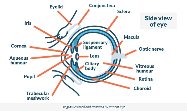
What is the vitreous humour?
Zurück zum InhaltThe vitreous humour is a clear jelly-like substance that makes up about 80% of the volume of the globe of the eye. It supports the shape of the globe of the eye whilst letting light through.
It is made up mainly of water but with some collagen and hyaluronic acid. The outside is made of fine fibres which are attached to the retina at the back and to the back of the lens at the front.
Side View of the Eye
Lesen Sie unten weiter
What are the causes of vitreous haemorrhage?
Zurück zum InhaltThe most common causes, accounting for about 90% of all cases of vitreous haemorrhage, are:
Bleeding from abnormal new blood vessels forming in advanced diabetic eye disease.
Bleeding from tears in the retina caused by vitreous detachment (see below).
Trauma to the eye (the most common cause in younger people).
Bleeding inside the eye can come from:
Abnormal blood vessels which grow because the back of the eye is short of oxygen. These are fragile and bleed easily. Conditions in which this can occur include:
Diabetic eye disease (die häufigste Ursache).
Retinopathy with Sichelzellenanämie.
Damage to the back of the eye in very premature babies who have been on oxygen in special care baby units.
Normal blood vessels which are damaged. They may be damaged by:
Posterior vitreous detachment, often because it causes a retinal tear (see below).
Retinal macroaneurysms - swollen blood vessels on the retina, usually related to Bluthochdruck, Arteriosklerose und rauchen.
Blunt trauma - suddenly compressing the eye - for example, if hit by a squash ball.
Penetrating trauma - this will cause bleeding throughout the eye. Penetrating trauma can occur from high-velocity injuries such as grinding and hammering. They do not always cause severe eye pain.
Subarachnoidalblutung, which can increase the pressure in the veins in the retina, causing them to bleed.
Surgery to the eye, particularly if it involves the inside of the eye.
Blood from behind the retina tracking through into the eye. This is the least common cause of vitreous haemorrhage. It may result from:
Tumours of the back of the eye (eye tumours are rare. The most common type is ocular melanoma).
Fragile new blood vessels behind the retina.
Posterior vitreous detachment is usually experienced between the ages of 60 and 80 - it happens to most of us between those ages. It occurs when the vitreous pulls away from the retina at the back. This can happen quite suddenly as the vitreous tends to shrink with age.
Most commonly there are no symptoms. Sometimes posterior vitreous detachment causes bleeding from the retina as it pulls away. Sometimes the retina is torn as the vitreous pulls away from it - in this case bleeding is more likely.
Most people experience posterior vitreous detachment in their 60s or later, and most do not have significant vitreous haemorrhage when it happens. However, episodes of noticeable floaters are very common and are probably caused by tiny bleeds.
Vitreous haemorrhage symptoms
Zurück zum InhaltThe symptoms of smaller bleeds (most bleeds are smaller bleeds) include:
Floaters in vision.
Cobweb haze.
Haze and shadows in the eye.
Red tint to the vision.
Symptoms most commonly affect one eye only, although both eyes can be affected.
Some people find that their vision is worse in the morning, because blood settles to the back of the eye overnight whilst lying down.
More severe bleeds cause:
Haziness of vision.
Blind spots or dark streaks.
The most severe bleeds cause visual loss, which can be complete, leaving the vision hazily red or black. For most people this is extremely alarming, particularly as it tends to come on very quickly with no clear explanation.
Lesen Sie unten weiter
How common is vitreous haemorrhage?
Zurück zum InhaltVitreous haemorrhage affects about 7 per 100,000 people each year. This makes it one of the most common causes of sudden deterioration in vision. It most often affects only one eye.
Who is likely to experience vitreous haemorrhage?
Zurück zum InhaltThe most common cause of vitreous haemorrhage is severe diabetic eye disease, which is mainly seen in older adults. The other common causes of vitreous haemorrhage also tend to occur in adults aged 60 and above, except for eye trauma, which can occur at any age.
How is vitreous haemorrhage diagnosed?
Zurück zum InhaltAn examination using a slit lamp is performed by opticians and eye specialists (ophthalmologists) to look in detail at the inside of the eye. The slit lamp will allow the examiner to see if there is blood in the vitreous.
Finding the source of the bleeding may be possible with the slit lamp, although if there is a lot of blood in the vitreous humour this prevents a clear view and it may therefore be difficult to know what has happened. In this case an ultrasound scan of your eye can help. Ultrasound can detect many causes of vitreous haemorrhage, including posterior vitreous detachment, retinal tears and detachments, tumours and foreign objects.
Sometimes an angiogram is needed. This test shows up the blood vessels in the back of the eye. This can be helpful if looking for abnormal blood vessels such as in diabetes.
Computerised tomography (CT) scanning of the eyes is useful if there is a suspicion of a penetrating injury.
What is the treatment for vitreous haemorrhage?
Zurück zum InhaltThe treatment of vitreous haemorrhage varies with the cause. Aims of treatment are to:
Find the source of the bleeding.
Stop the bleeding.
Repair any damage to the retina before it results in permanent loss of vision.
Restore normal vision.
Once the source of the bleeding has been identified, treatment will depend on the cause. If there is not too much blood in the vitreous and the source of bleeding can be seen then it is treated. This means laser treatment to bleeding vessels and any other abnormal vessels, and repair to any tears in the retina. After this it is a matter of waiting for the blood to slowly clear as the red blood cells break down. This can take several weeks.
Strenuous activity should be avoided for several days at least, as this might dislodge clots and trigger new bleeding. It is also advised to sleep with the head of the bed elevated, as this allows the blood in the vitreous to settle into the bottom of the eye, out of the line of vision.
If the blood in the vitreous obscures the view and prevents treatment of the bleeding then the entire vitreous may be removed first. This procedure is called a vitrectomy. Doctors will perform a vitrectomy if they can't see the back of the eye, or if the view isn't good enough to treat the bleeding there safely.
Specific treatments
Laserphotokoagulation
This is the usual treatment for fragile abnormal vessels. Treating them both stops the bleeding and prevents later bleeding. Laser photocoagulation is also used in repairing damage to the retina, including retinal detachments.
Anti-VEGF injections
Anti-VEGF injections (a type of intravitreal injection) aim to shrink abnormal new vessels which have formed in the eye. They are sometimes used in patients with diabetes, in addition to other treatments like laser photocoagulation and vitrectomy, in order to reduce bleeding.
Kryotherapie
This is also used as a treatment for retinal tears and retinal detachments.
Vitrectomy
Vitrectomy is removal of the vitreous humour completely, together with the membrane that surrounds it. This is done when there is so much blood in the vitreous that it is impossible to diagnose and treat the cause. Vitrectomy is also sometimes performed if the blood in the vitreous is clearing very slowly and vision remains impaired.
Waiting
Waiting is commonly the chosen option once the bleeding has stopped. Most cases of vitreous haemorrhage do not require vitrectomy. The blood clears slowly from the vitreous, allowing light to pass through it again. If the underlying vision is undamaged then normal vision will be restored.
Allgemeine Maßnahmen
Anyone with a suspected vitreous haemorrhage will usually be seen by an eye specialist on the same day. This is because sudden loss of vision is considered an eye emergency. The aim is to ensure accurate diagnosis and to avoid permanent loss of vision which could occur if there is a Netzhautablösung behind the bleeding.
Anyone who has a sudden loss of vision or any other unexplained disturbance in vision in one or both eyes should:
See a GP or optician, or attend an accident and emergency department, as soon as possible.
Try to remain calm - most vitreous haemorrhages respond well to treatment and there is a very good chance that vision will return to where it was.
Rest for a few hours a day sitting upwards and elevate the head at night on pillows.
Ask a GP to check blood pressure - raised blood pressure increases the chance of further bleeding.
Avoid any heavy lifting, which can increase the chance of further bleeding.
What is the outlook for vitreous haemorrhage?
Zurück zum InhaltThe outlook (prognosis) in vitreous haemorrhage depends both on the cause and on the severity.
Vitreous haemorrhage resulting from posterior vitreous detachment usually has a good prognosis, with restoration of vision, particularly if the eye is otherwise normal.
Where severe diabetic eye disease or macular degeneration has resulted in abnormal blood vessels, the outlook for the vision is much less good. The outlook in penetrating eye injury is often poor.
How is vitreous haemorrhage prevented?
Zurück zum InhaltPrevention of vitreous haemorrhage involves preventing the underlying causes. This includes careful and regular management of diabetic eye disease (which tends to be worse in less well-controlled diabetes) and high blood pressure, and giving up smoking.
The eye should always be protected from trauma during high-risk activities such as filing, grinding and hammering, using firearms and playing sports with high-speed balls, such as squash.
Dr. Mary Lowth ist eine Autorin oder die ursprüngliche Autorin dieses Merkblatts.
Patient picks for Augenkrankheiten

Augengesundheit
Netzhautvenenverschluss
Die Netzhautvenenverschluss tritt auf, wenn eine der kleinen Venen in der Netzhaut durch ein Blutgerinnsel blockiert wird.
von Dr. Toni Hazell, MRCGP
Augengesundheit
Akutes Winkelblockglaukom
Acute angle-closure glaucoma is a serious eye condition that occurs when the fluid pressure inside the eye rises quickly, leading to sudden, severe eye pain, a red eye and reduced or blurred vision. It is a medical emergency.
von Dr. Philippa Vincent, MRCGP
Häufig gestellte Fragen
Can vitreous haemorrhage affect both eyes, or just one?
Vitreous haemorrhage most commonly affects only one eye. However, it is possible for both eyes to be affected.
Why might my vision be worse in the morning if I have a vitreous haemorrhage?
If you have a vitreous haemorrhage, your vision might be worse in the morning because blood settles to the back of the eye overnight while you are lying down.
Are there any immediate steps I should take if I experience sudden vision loss?
If you experience sudden loss of vision or any other unexplained vision disturbance in one or both eyes, you should see a GP, optician, or attend an accident and emergency department as soon as possible. It's also advised to try and remain calm, rest for a few hours a day sitting upwards, and elevate your head at night on pillows. You should also avoid any heavy lifting.
What is the purpose of an ultrasound scan if a lot of blood is present in the eye?
If there is a lot of blood in the vitreous humour, it can obstruct the view during a routine examination, making it difficult to identify the cause of the haemorrhage. In such cases, an ultrasound scan of your eye can help. It can detect various causes, including posterior vitreous detachment, retinal tears and detachments, tumours, and foreign objects.
How long does it typically take for the blood to clear after a vitreous haemorrhage?
After the source of bleeding has been treated, if there wasn't too much blood and the source could be seen, it's a matter of waiting for the blood to slowly clear as the red blood cells break down. This process can take several weeks.
Why is it important to prevent underlying conditions like diabetes and high blood pressure to avoid vitreous haemorrhage?
Preventing vitreous haemorrhage involves managing its underlying causes. Careful and regular management of diabetic eye disease, which tends to be worse in less well-controlled diabetes, and high blood pressure are crucial steps. This proactive approach helps reduce the risk of abnormal or fragile blood vessels forming that can lead to bleeding.
Weiterführende Literatur und Referenzen
- Shaikh N, Srishti R, Khanum A, et al; Vitreous hemorrhage - Causes, diagnosis, and management. Indian J Ophthalmol. 2023 Jan;71(1):28-38. doi: 10.4103/ijo.IJO_928_22.
- Mathen,T, Leng, T, Shah VA; Terson Syndrome, EyeWiki - American Academy of Ophthalmology
Lesen Sie unten weiter
About the authorView full bio

Dr Colin Tidy, MRCGP
Allgemeinmediziner, Medizinischer Autor
MBBS, MRCGP, MRCP (Paediatrics), DCH
Dr. Colin Tidy ist ein NHS-Arzt mit Sitz in Oxfordshire.
About the reviewerView full bio

Dr Doug McKechnie, MRCGP
Medizinischer Autor
MA, MBBS, MSc, DRCOG, MRCP(UK), MRCGP(2021), FHEA
Dr. Doug McKechnie ist ein NHS-Hausarzt, der in London arbeitet. Er arbeitet klinisch in Vollzeit und ist außerdem stellvertretender Leiter des Moduls für klinische und berufliche Praxis an der University College London Medical School.
Artikelverlauf
Die Informationen auf dieser Seite wurden von qualifizierten Klinikern verfasst und begutachtet.
Next review due: 28 Jun 2027
29 Jun 2024 | Neueste Version

Fragen, teilen, verbinden.
Durchsuchen Sie Diskussionen, stellen Sie Fragen und teilen Sie Erfahrungen zu Hunderten von Gesundheitsthemen.

Fühlen Sie sich unwohl?
Bewerten Sie Ihre Symptome online kostenlos
Abonnieren Sie den Patienten-Newsletter
Ihre wöchentliche Dosis klarer, vertrauenswürdiger Gesundheitsberatung - geschrieben, um Ihnen zu helfen, sich informiert, selbstbewusst und in Kontrolle zu fühlen.
By subscribing you accept our Datenschutzrichtlinie. Sie können sich jederzeit abmelden. Wir verkaufen Ihre Daten niemals.