Gastroskopie
Endoskopie
Begutachtet von Dr Hayley Willacy, FRCGP Zuletzt aktualisiert von Dr Philippa Vincent, MRCGPLast updated 3. Juli 2024
Erfüllt die Anforderungen des Patienten Richtlinien des Patienten
- HerunterladenHerunterladen
- Teilen
- Language
- Diskussion
- Audio-Version
- Add to preferred sources on Google
In dieser Serie:VerdauungsstörungenGastritisFunktionelle DyspepsieMagengeschwürZwölffingerdarmgeschwürHelicobacter pylori
Gastroscopy is a test to look inside the gullet (oesophagus), the stomach and the first part of the gut (small intestine) known as the duodenum.
Hinweis: the information below is a general guide only. The arrangements, and the way tests are performed, may vary between different hospitals and areas. Always follow the instructions given by your doctor or local hospital.
At a glance
A gastroscopy is a procedure to look inside your oesophagus, stomach, and duodenum using a thin, flexible tube with a camera.
It is used to investigate symptoms like recurring indigestion, heartburn, tummy pain, repeated vomiting, or difficulty swallowing.
The procedure usually takes about 10 minutes and is often done as a day case.
You might be given a local anaesthetic spray or lozenge for your throat, and a sedative to help you relax.
After the procedure, if you've had a sedative, avoid driving, operating machinery, or drinking alcohol for 24 hours.
Seek immediate medical help if you experience worsening tummy pain, fever, difficulty breathing, or vomiting blood within 48 hours.
In diesem Artikel:
Video picks for Endoskopie
Lesen Sie unten weiter
What is a gastroscopy?
Gastroskopie
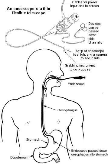
A gastroscopy is a test where an operator - usually a doctor or nurse - looks into the upper part of the gut (the upper gastrointestinal tract) with a camera.
The upper gut consists of the gullet (oesophagus), the stomach and the first part of the gut (small intestine) known as the duodenum. The operator uses an endoscope to look inside the gut.
An endoscope is a thin, flexible telescope. It is about as thick as a little finger. The endoscope is passed through the mouth, down the food pipe into the oesophagus and down towards the stomach and duodenum.
The tip of the endoscope contains a light and a tiny video camera so the operator can see inside the gut.
The endoscope also has a side channel down which various instruments can pass. These can be manipulated by the operator. For example, the operator may take a small sample (Biopsie) from the inside lining of the stomach by using a thin 'grabbing' instrument which is passed down a side channel.
The procedure is sometimes called an "upper GI endoscopy" - this refers to the upper gastro-intestinal tract.
Why do I need a gastroscopy?
Zurück zum InhaltA gastroscopy may be advised for symptoms such as:
Repeated (recurring) indigestion.
Recurring heartburn.
Pains in the upper tummy (abdomen).
Repeatedly being sick (vomiting).
Schwierigkeiten beim Schlucken.
Other symptoms thought to be coming from the upper gut, stomach or oesophagus.
Lesen Sie unten weiter
What is a gastroscopy looking for?
Zurück zum InhaltInflammation of the gullet (oesophagus), called oesophagitis. The operator will see areas of redness on the lining of the oesophagus.
Magengeschwür und Zwölffingerdarmgeschwür. An ulcer looks like a small, red crater on the inside lining of the stomach or on the first part of the gut (small intestine) known as the duodenum.
Inflammation of the duodenum (duodenitis) and inflammation of the stomach (Gastritis).
Stomach und Speiseröhrenkrebs cancer.
Hiatushernie
Various other rare conditions.
What happens during a gastroscopy?
Zurück zum InhaltGastroscopy is usually done as an outpatient 'day case'. It is a routine test which is commonly done. Before the test the operator will explain what is going to happen and ask for a consent form to be signed.
The operator may numb the back of the throat by spraying on some local anaesthetic or giving an anaesthetic lozenge to suck. A sedative may also be given to help relaxation. This is usually given by an injection into a vein in the back of the hand. The sedative causes drowsiness but is not a general anaesthetic and does not put someone to sleep.
You lie on your side on a couch. You are asked to put a plastic mouth guard between your teeth. This works to protect your teeth and stops you biting the endoscope. The operator will then ask you to swallow the first section of the endoscope. Modern endoscopes are quite thin but some people may find this difficult.
The operator then gently pushes it further down the gullet (oesophagus) and into the stomach and the first part of the gut (small intestine) known as the duodenum. The video camera at the tip of the endoscope sends pictures to a screen.
The operator watches the screen for abnormalities of the oesophagus, stomach and duodenum. Air is passed down a channel in the endoscope into the stomach to make the stomach lining easier to see. This may cause a bloating sensation and belching.
The operator may take one or more small tissue samples (biopsies) of parts of the inside lining of the gut - depending on why the test is done and what they see on the screen. This is painless. The biopsy samples are sent to the laboratory for testing and to look at under the microscope. The endoscope is then gently pulled out.
How long does a gastroscopy take?
A gastroscopy usually takes about 10 minutes. However, at least two hours should be allowed for the whole appointment. This is to prepare, give time for the sedative to work, for the gastroscopy itself and to recover. A gastroscopy may be quite uncomfortable but it does not usually hurt.
Lesen Sie unten weiter
How do you prepare for a gastroscopy?
Zurück zum InhaltYou should receive instructions from the hospital department before your test. The sort of instructions given commonly include:
Avoid eating or drinking for 4-6 hours before the test. The stomach needs to be empty. (Small sips of water may be allowed up to two hours before the test.)
If you have a sedative you will need somebody to accompany you home.
Advice about medication which may need to be stopped before the test.
What can I expect after a gastroscopy?
Zurück zum InhaltMost people are ready to go home after resting for half an hour or so.
Wenn Sie ein Beruhigungsmittel eingenommen haben - you may take a bit longer to be ready to go home. The sedative will normally make you feel quite pleasant and relaxed. However, you should not drive, operate machinery or drink alcohol for 24 hours after having the sedative. You will need somebody to accompany you home and to stay with you for 24 hours until the effects have fully worn off. Most people are able to resume normal activities after 24 hours.
The operator writes a report and sends it to the doctor who requested the gastroscopy, usually a gastroenterologist but occasionally another specialist or GP. The result from any sample (biopsy) may take some days or even weeks, which can delay the report being sent. The operator may inform you about what they saw before you leave. However, if you have had a sedative you may not remember afterwards what you were told. Therefore, you may wish to have a relative or close friend with you who may be able to help remember what was said.
Is gastroscopy reliable?
Zurück zum InhaltGastroscopy is a good test for seeing abnormalities in the upper gut. However, it is not fool-proof. For example, gastroscopy may not detect a small number of cases of early ulcers or early cancer. Sometimes a repeat gastroscopy may be advised if symptoms persist or become worse, even if a previous gastroscopy was reported as normal.
Is a gastroscopy dangerous?
Zurück zum InhaltMost gastroscopies are done without any problem. Some people have a mildly sore throat for a day or so afterwards. You may feel tired or sleepy for several hours if you have a sedative. There is a slight risk of developing a chest infection or pneumonia following a gastroscopy.
Occasionally, the endoscope causes some damage to the gut. This may cause bleeding, infection and rarely a hole (perforation). If any of the following occur within 48 hours after a gastroscopy, seek immediate medical help:
Bauchschmerzen (abdominale Schmerzen). (In particular, if it becomes gradually worse, and is different from or more intense than any 'usual' indigestion pains or heartburn.)
Erhöhte Temperatur (Fieber).
Atembeschwerden.
Blut erbrechen.
A small number of people have a Herzinfarkt oder Schlaganfall during, or soon after, a gastroscopy. These tend to be older people who are already in poor health. These serious complications are rare in most people who are otherwise reasonably healthy.
Rarely, some people have an allergic reaction to the sedation.
Patient picks for Endoskopie

Chirurgie und Verfahren
Nasoendoskopie
Eine Nasoskopie ist ein Test, um das Innere der Nase (Nasengang), den hinteren Teil des Rachens (Pharynx) und den Kehlkopf (Larynx) zu untersuchen. Manchmal wird sie auch als flexible Nasenendoskopie oder FNE bezeichnet. Hinweis: Die nachstehenden Informationen sind nur eine allgemeine Orientierung. Die Abläufe und die Durchführung der Tests können in verschiedenen Krankenhäusern variieren. Befolgen Sie stets die Anweisungen Ihres Arztes oder des örtlichen Krankenhauses.
von Dr. Doug McKechnie, MRCGP

Chirurgie und Verfahren
Sigmoidoskopie
A sigmoidoscopy is a test that looks at the rectum and lower part of the large intestine. Note: the information below is a general guide only. The arrangements and the way tests are performed, may vary between different hospitals. Always follow the instructions given by your doctor or local hospital.
by Dr Claudia Berty, MRCGP
Häufig gestellte Fragen
What is the difference between a gastroscopy and an upper GI endoscopy?
There is no difference; they are two terms for the same procedure. Both refer to looking into the upper gastrointestinal tract (gullet, stomach, and the first part of the small intestine) using an endoscope.
Will I feel any pain if a biopsy is taken during the procedure?
No, taking a small tissue sample (biopsy) from the lining of the gut during a gastroscopy is described as painless. These samples are then sent to a laboratory for further testing.
Why might I feel bloated or belch during the gastroscopy?
During the procedure, air is passed down a channel in the endoscope into your stomach. This is done to make the stomach lining easier for the operator to see, and it can cause a bloating sensation and belching.
Can I drive myself home after a gastroscopy?
If you have been given a sedative, you must not drive for 24 hours after the procedure. You will need someone to accompany you home and stay with you for 24 hours until the effects of the sedative have fully worn off.
What should I do if I feel worse after the gastroscopy?
You should seek immediate medical help if you experience worsening tummy pain, a raised temperature, difficulty breathing, or vomit blood within 48 hours after the procedure. These could be signs of a rare but serious complication.
Will I get my results immediately after the gastroscopy?
The operator may inform you about what they observed before you leave. However, if you had a sedative, you might not remember what was said. The results from any biopsy samples can take several days or even weeks to come back, which will delay the final report being sent to your doctor.
Can a gastroscopy miss any problems?
While gastroscopy is effective for detecting abnormalities, it's not entirely foolproof. It may occasionally miss early ulcers or early cancers. If your symptoms continue or worsen, a repeat gastroscopy might be recommended even if a previous one was clear.
Weiterführende Literatur und Referenzen
Lesen Sie unten weiter
About the authorView full bio

Dr Philippa Vincent, MRCGP
Allgemeinmediziner, Medizinischer Autor
MB BS, Bsc, MRCGP (2000), DCH, DFSRH, DRCOG
Dr Philippa Vincent is an NHS GP working in North London.
About the reviewerView full bio

Dr Hayley Willacy, FRCGP
Allgemeinmediziner, Medizinischer Autor
MBChB (1992), DRCOG, DFFP, MRCOG (Part 1) MRCGP (2007), DFSRH (2013), MSc - medical education (2020)
Dr Hayley Willacy was an NHS GP working in northwest England, who retired from clinical practice in 2022 after 30 years.
Artikelverlauf
Die Informationen auf dieser Seite wurden von qualifizierten Klinikern verfasst und begutachtet.
Nächste Überprüfung fällig: 2. Juli 2027
3. Juli 2024 | Neueste Version

Fragen, teilen, verbinden.
Durchsuchen Sie Diskussionen, stellen Sie Fragen und teilen Sie Erfahrungen zu Hunderten von Gesundheitsthemen.

Fühlen Sie sich unwohl?
Bewerten Sie Ihre Symptome online kostenlos
Abonnieren Sie den Patienten-Newsletter
Ihre wöchentliche Dosis klarer, vertrauenswürdiger Gesundheitsberatung - geschrieben, um Ihnen zu helfen, sich informiert, selbstbewusst und in Kontrolle zu fühlen.
By subscribing you accept our Datenschutzrichtlinie. Sie können sich jederzeit abmelden. Wir verkaufen Ihre Daten niemals.