Ascites
Peer reviewed by Dr Toni Hazell, FRCGPLast updated by Dr Caroline Wiggins, MRCGP Last updated 20 Jun 2026
Meets Patient’s editorial guidelines
- DownloadDownload
- Share
- Language
- Discussion
- Audio Version
- Add to preferred sources on Google
Medical Professionals
Professional Reference articles are designed for health professionals to use. They are written by UK doctors and based on research evidence, UK and European Guidelines. You may find the Cirrhosis article more useful, or one of our other health articles.
What is ascites?
Ascites is the excessive accumulation of fluid in the abdominal cavity. For ascites fluid to be detectable by clinical examination there has to be at least 1500 ml present (slightly less in a small, thin person, but significantly more in a person with obesity). Ultrasound can detect much smaller volumes (≤500 ml).
Ascites that is not infected and not associated with hepato-renal syndrome may be graded as follows:1
Grade 1 is mild ascites and is only detectable by ultrasound examination.
Grade 2 is moderate ascites causing moderate symmetrical distension of the abdomen.
Grade 3 is large ascites causing marked abdominal distension.
Refractory ascites can be divided into two groups:
Diuretic-resistant ascites is refractory to dietary sodium restriction and intensive diuretic treatment for at least one week.
Diuretic-intractable ascites is refractory to therapy due to the development of diuretic-induced complications that preclude the use of an effective dose of diuretic.
Causes of ascites1
Cirrhosis:
Approximately 55% of patients presenting with ascites have underlying alcohol-related cirrhosis.
Fluid retention (primarily ascites but also peripheral oedema and pleural effusions) is the most frequent complication of end-stage liver disease. It significantly impairs the quality of life of patients with cirrhosis and is associated with poor prognosis - one-year and five-year survival rates of 85% and 56% respectively.2
Beware the patient with a very long history of stable cirrhosis who then develops ascites - hepatocellular carcinoma must be excluded.
Malignancy accounts for around 30%. The usual causes are:
Malignancies of the gastrointestinal tract (carcinoma of stomach, colon, pancreas; primary hepatocellular carcinoma and metastatic liver cancer).
Carcinoma of ovary.
Hodgkin's lymphoma and non-Hodgkin's lymphoma.
Metastatic carcinoma within the abdominal cavity.
Heart failure.
Nephrotic syndrome.
Protein-losing enteropathy.
Tuberculosis.
Pancreatitis.
Other rare causes, including hypothyroidism.
Iatrogenic - eg, ovarian hyperstimulation as a consequence of IVF procedures.
Ascites symptoms (presentation)
Abdominal distension.
Weight gain as a result of water retention.
Discomfort: tense ascites is very uncomfortable but prior to this stage there is simply abdominal distension with only very mild discomfort. Malignancy-related ascites is frequently painful.
Nausea and appetite suppression: tense ascites presses on the stomach and bowel.
Increasing dyspnoea: due to limited venous return from the lower limbs (pressure on the inferior vena cava) and impaired expansion of the lungs (pressure on the diaphragm).
There may be other symptoms related to the cause of the ascites.
Examination
Perform a full abdominal examination.
Hepatic failure - ascites
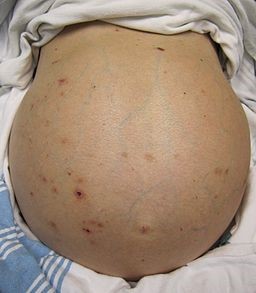
© James Heilman, MD, CC BY-SA 3.0, via Wikimedia Commons
Look at the patient, both with them lying down and standing up. The shape of the abdomen often suggests ascites fluid. On lying down, the flanks are full but on standing the ascites fluid accumulates in the lower abdomen.
The high intra-abdominal pressure may push out an umbilical hernia or even an inguinal hernia.
There may be stigmata of other diseases or the cause of the ascites. Look for signs of liver disease and cirrhosis, including:
Jaundice.
Muscle wasting.
Gynaecomastia.
Spider naevi.
Palmar erythema.
Rarely, a firm nodule in the umbilicus (known as Sister Mary Joseph's nodule) is found and suggests peritoneal carcinomatosis originating from gastric, pancreatic, or hepatic primaries. A left-sided supraclavicular node, or Virchow's node, suggests the presence of upper abdominal malignancy.
Examination for ascites
Shifting dullness is used to detect ascites. One study found the absence of flank dullness to be the most accurate predictor against the presence of ascites - the probability of ascites without flank dullness was less than 10% :
Percuss from the level of the umbilicus and repeat moving laterally towards one side.
When the sound becomes dull, keep your fingers there to mark the spot and ask the patient to move on to the opposite side.
Wait briefly for the fluid to sink and then percuss again. If it is now resonant, that is a positive sign. Percuss down until dullness is reached again.
Repeat on the other side.
False positives do occur, probably from dilated coils of small intestine reacting to gravity.
At least 1500 ml of fluid must be present for a result. An ultrasound scan will detect much less fluid with greater certainty.
Large ascites can be detected by a 'fluid thrill'. This test requires two examiners. One person places the side of the palm of one hand firmly on the centre of the abdomen, with the fingers pointing towards the groin. The second person places the palm of one hand on one flank and then flicks the other flank. In large ascites, a palpable fluid thrill can be felt by the palm resting on the opposite flank.
Monitoring
Simple assessment of the progress of ascites may be made by:
Serial measurements of the abdominal girth - ensure the tape measure is placed in the same position each time.
Serial measurement of weight - rapid changes indicate fluid gain or loss which are much faster than gain or loss of fat or lean body mass.
Differential diagnosis
The differential diagnosis of ascites is with other causes of abdominal mass, especially large cysts, although sometimes plain obesity may seem like ascites. The essential feature is the fluidity and shifting with position.
Investigations1
The cause of the ascites is often apparent after an adequate history and examination. The aims of investigation for ascites are:
Confirming the presence of ascites.
Finding the cause for the ascites.
Assessing for complications due to the ascites.
Initial investigations
Blood tests to look for the cause and for complications.
Imaging. Abdominal ultrasound is a very sensitive way of assessing ascites and may also show the causative pathology. CXR may show pleural effusion, evidence of pulmonary metastases or heart failure. If ultrasound has failed to reveal a cause then CT scanning may be used.
Diagnostic paracentesis is recommended in all patients with new-onset ascites. See the separate Ascites tapping article.
Ascites treatment and management1
Treatment of the underlying cause.
Management of the ascites varies depending on the cause. See below for details.
Salt restriction
Patients with ascites due to cirrhosis should have a moderately salt-restricted diet with daily salt intake of no more than 5-6.5 g (87 mmol-113 mmol sodium). In reality, this means a no added salt diet with avoidance of precooked meals.
Drugs
Along with salt restriction, ascites due to cirrhosis is managed by diuretic therapy:
Diuretics:
Spironolactone is the initial choice in cirrhosis: it increases sodium excretion and potassium reabsorption in the distal tubules. 100 mg/day can gradually be increased to 400 mg as necessary. Serum potassium levels need monitoring, as hyperkalaemia frequently limits spironolactone's use.
Loop diuretics are used as an adjunct to spironolactone. Start cautiously with, for example, furosemide 40 mg/day, although up to 160 mg/day may be used. Again, monitoring is required to assess for electrolyte disturbance, particularly hyponatraemia.
Amiloride may be used in patients intolerant of spironolactone but it is not as effective.
Midodrine:
Portal hypertension and splanchnic vasodilatation are major factors in the development of ascites. Vasopressors such as midodrine, an α-adrenergic agonist, have been used in non-azotemic patients with ascites. This results in significant increase in mean arterial pressure and urine sodium excretion and significant decreases in plasma renin and aldosterone.
The British Society of Gastroenterology (BSG) guidelines suggest that it should be considered for patients with refractory ascites on a case-by-case basis. This is an off-licence use and would be initiated by secondary care.
Diuretics are not commonly used to manage malignant ascites.3
Anti-neoplastic therapy for malignant ascites.3
Therapeutic paracentesis
Patients with large or refractory ascites generally benefit from therapeutic paracentesis. This needs to be a sterile procedure and is undertaken in hospital along with an infusion of human albumin solution.
Surgical
A transjugular intrahepatic portosystemic shunt (TIPSS) may be considered for patients with refractory ascites though there are a number of contraindications.
Palliative care1
The only curative option for untreatable ascites is liver transplantation. If the patient is not suitable for liver transplantation, the focus is then on the control of symptoms. The most common palliative treatment is repeated ascitic drainage in hospital. Alternative treatments for untreatable ascites, such as indwelling drains may be considered in malignant ascites.
Ascites complications
Electrolyte disturbance by diuretics.1
Spontaneous bacterial peritonitis (SBP) - see also the separate Intra-abdominal sepsis and abscesses article:
This occurs in 10-30% of patients with ascites and has a mortality rate of 20%.
It is frequently asymptomatic but most will have some symptom(s) such as fever, mild abdominal pain, vomiting or confusion.
Suspect SBP where patients present with hepatic encephalopathy, renal impairment or peripheral leukocytosis without any obvious precipitating factor.
Immediate empirical antibiotic therapy should be commenced.
Prophylactic antibiotics for SBP should be given in certain patient populations.
Prognosis
Patients with cirrhosis who develop ascites have a one-year mortality rate of 15% and a five-year survival rate of 44%.5
Malignancy ascites tends to suggest widespread disease and a poor prognosis.
Exclusive updates for healthcare professionals
Stay informed with the latest clinical updates, professional insights, and evidence-based guidance. The Patient Pro newsletter curates essential content for healthcare professionals—delivered straight to your inbox.
By subscribing you accept our Privacy Policy. You can unsubscribe at any time. We never sell your data.
Further reading and references
- Sundaram V, Manne V, Al-Osaimi AM; Ascites and spontaneous bacterial peritonitis: recommendations from two United States centers. Saudi J Gastroenterol. 2014 Sep-Oct;20(5):279-87. doi: 10.4103/1319-3767.141686.
- Aithal GP, Palaniyappan N, China L, et al; Guidelines on the management of ascites in cirrhosis. Gut. 2021 Jan;70(1):9-29. doi: 10.1136/gutjnl-2020-321790. Epub 2020 Oct 16.
- Kashani A, Landaverde C, Medici V, et al; Fluid retention in cirrhosis: pathophysiology and management. QJM. 2008 Feb;101(2):71-85. Epub 2008 Jan 9.
- Management of ascites in ovarian cancer patients. Scientific impact paper No 45. November 2014.
- Lenz K, Buder R, Kapun L, et al; Treatment and management of ascites and hepatorenal syndrome: an update. Therap Adv Gastroenterol. 2015 Mar;8(2):83-100. doi: 10.1177/1756283X14564673.
- Biecker E; Diagnosis and therapy of ascites in liver cirrhosis. World J Gastroenterol. 2011 Mar 14;17(10):1237-48. doi: 10.3748/wjg.v17.i10.1237.
About the authorView full bio

Dr Caroline Wiggins, MRCGP
General Practitioner, Medical Author
MBBS Honours (with Distinction), MRCGP (2016), MSc.SEM (with Distinction), BSc (Hons)
Dr Caroline Wiggins is a GP locum currently in the South-West of England.
About the reviewerView full bio

Dr Toni Hazell, FRCGP
MBBS, BSc, FRCGP, DFSRH, Dip GU med, DRCOG, DCH (London, UK, 2000)
Dr. Toni Hazell qualified from St. Mary’s Hospital Medical School and did her VTS at Northwick Park Hospital.
Article history
The information on this page is written and peer reviewed by qualified clinicians.
Article also available in English, German, Spanish, French, Italian, Portuguese, Hindi, Hebrew, Arabic, and Swedish.
Next review due: 20 Dec 2030
20 Jun 2026 | Latest version

Ask, share, connect.
Browse discussions, ask questions, and share experiences across hundreds of health topics.

Feeling unwell?
Assess your symptoms online for free
More in history and examination
- Antenatal examinations and diagnosis of pregnancy
- Back pain in children
- Blurred vision
- Cerebellar disorders
- Foster Kennedy's syndrome
- Gynaecological examination and history
- Hearing testing and screening in young children
- High-altitude illness
- History and physical examination
- Itching
- Lower urinary tract symptoms in men
- Lumps
- Marfan's syndrome
- Mild cognitive impairment
- Nasal discharge
- Neurological examination of the lower limbs
- Nocturnal enuresis in children
- Palmar erythema
- Post-inflammatory hyperpigmentation of skin
- Snoring